Sorry for mistakes,page on traslation working
Extensive multi-disciplinary research in many fields of application, have prompted me to investigate the biochemistry field ... and...
The cell represents LIFE in all its organizations.
Research, Progress, Technology, do not stop and thanks to researchers in the Universities , every day there are some fantastic discoveries in everything that is called LIFE.
My thoughts go out to these people, who bring great news in various fields of study.
Research often is underestimated by all of us, but it thanks to it that medicine, pharmacy and numerous disciplines lead to the cure of many diseases.
Pharmacy Science of Cosmetics
Pier
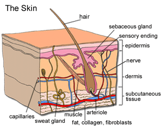
" THE SKIN "
Extended search
by Pier in Project Light Radiance Cosmetics Italy-France
The skin (or skin) is the outer covering of our body and, more generally, of a vertebrate. In mammals and humans in particular, is the largest organ of the integumentary system (its surface is about 2 square meters) and protects the underlying tissues (muscles, bones, internal organs). The skin consists of a series of ectodermal and mesodermal tissues, which can have various colors, organic and physiological structure, meeting even the aging process more or less visible.
Physiological role
As mediator between the body and the outside world, the skin performs different functions in vertebrates:
Security: as anatomical barrier against any potential pathogens and pests, is the first line of defense against external aggression. It also contains Langerhans cells, whose function is to present the antigen, which are part of the acquired immune system [1] [2], because the skin is the first line of defense against the action of potential pathogens [1].
Sensitivity: in the skin are numerous nerve endings that detect changes in temperature (thermoreceptors), pressures (pressoreceptors), vibration and painful sensations (algocettori), mean the sense of touch.
Control of evaporation: The skin is dry and relatively impermeable barrier against fluid loss, [2] also adjusting the excretion of electrolytes through perspiration.
Heat regulation: The skin has a far greater blood flow to those who are its actual metabolic needs, this feature make it an ideal medium for the regulation of body temperature. The vasodilation causes an increase of local blood flow, which favors the transfer of heat to the external environment, conversely, vasoconstriction, reducing the amount of blood in transit, keeps heat loss.While in humans are almost insignificant in animals hair erector muscles contribute by their contraction (gooseflesh) to produce heat, sweating as easily hand the loss of heat, and acts as a thermal insulator, as a regulator of body temperature and prevents excessive water loss [2].
Power Consumption: Since small amounts of oxygen, nitrogen and carbon dioxide can diffuse freely in the epidermis, some animals (especially small amphibians) will use their skin as a single organ respiratory function, contrary to what is usually believed, humans humans do not absorb oxygen through the skin [3].
defense / offense: different skin appendages (nails, horns in rhinoceros) serve as a means of defense or offense, the camouflage can be scored the same in both categories, depending on whether the prey is to use it (to escape predators) or predators (or attract prey to approach them without arousing suspicion)
Sexual attraction (pigmentation)
Reserve and the role of synthetic: it is a reservoir of lipids and water and allows the synthesis of certain substances such as vitamin D needed
Structure
The skin has a different structure in different classes of vertebrates (fishes, amphibians, reptiles, birds and mammals [4]). The skin of fishes and reptiles is characterized by the presence of protective scales, while the presence of bird feathers, the skin of amphibians but not a rigid barrier to the passage of chemical substances, so much so that it is often subject to phenomenaosmotic. The skin of mammals is made unique by the presence of hairs, even in marine mammals, which also appear hairless. A sufficient density pilifera results in fur, which have an additional thermal insulation properties but can also serve as a secondary sexual character or mimetic character. The skin of some animal characteristics such as thickness and stiffness make it usable to produce leather.
Aging
As the skin ages, it becomes increasingly thin and fragile, due to the fact that cell regeneration slows and passes from the normal 3-4 weeks at 4 or even 6 weeks. This is due to the thinning of the thyroid hormones that regulate the operation. Wrinkles are a result of the decrease in elasticity of the skin, and not just aging. Indeed, it is possible to find wrinkles in the very young, this is due to the fact that wrinkles are formed in which there are major muscle movements, which require special skin elasticity.
The skin surface is not uniform, has a design very complex and varies from area to area for the presence of parallel surface grooves that determine, especially in the fingertips of figures features (dermatoglyphics), which vary from individual to individual. On the palms and soles of the feet are also deep grooves, while in areas under joint movement are skin folds, the wrinkles that form on the face and neck of the individuals after a certain age are due to loss of elasticity of skin.
Pigmentation / coloration
The color of the skin in humans depends on many variables (thickness of the stratum corneum, sanguificazione status, presence and optical absorption of melanin, carotene, hemoglobin). Therefore, the color varies not only between the various ethnic groups, but also from individual to individual and also varies even slightly in different parts of the body of the same individual.
The color pigment that is due to special cells called chromatophores, melanophores these may be present in both the dermis into the epidermis, or xantofori iridofori in the dermis.
The color of the skin depends mainly on melanin, which is more and more concentrated to give a dark skin, this color is stimulated during the warm period, giving it more color and protect the skin from aging.
Once past the warm season or tanned skin can gradually remove themselves so adherent, forming the cuticle.
Hygiene
It is common in modern human societies frequent cleaning of the skin, aimed primarily at maintaining a certain level of hygiene, since a lack of cleanliness and promotes the spread of pathogens and diseases. This practice helps the skin to perform its functions and retain the coating, protection, secretion, thermoregulation, sensory and immune response, but it is obvious that even without such care the skin is still able to perform their duties with excellence.With the recent development of a complex society and the need for stronger contact between individuals, even far beyond the distances of intimacy with each other, has emerged the need to mitigate and reduce the body odor in a more rigorous rules and practice of hygiene more stringent than they did in the past, so the two annual washing with soap is passed to the Renaissance practice more intense and delicate.
Must be remembered that the washing of the epidermis (skin) with detergents, if practiced intensely aggressive and has an exfoliating effect, so compared to a removal of dirt you can have to remove the natural protective layer, such as the surface layer of sebum that keeps the skin soft and the condition of being soft. The heavy use of soaps or detergents can then produce a temporary state of decreased skin's own protection systems and a worsening of the state itself, which is normal after a few hours. In such a state can be addressed by natural or artificial products details.
The increased use of common materials or accessories of synthetic detergents (non-natural), has placed the need to clarify the effects of the substances used.
In the past, over the centuries, when the development has imposed thickening civil population, companies have often developed urban structures, and related social behavior, and social health of great importance for that part of the body that ultimately is the layer of contiguous with the outside world and with other individuals. In this sense, the Roman baths are located, or Japanese, in which the care and stimulation of this "frontier" of contact resulted in complex social rituals, and behavioral (use of ointments, massage practice, etc..) Even not strictly related to only appearance and hygiene.
Mechanical properties
As the soft tissue, the skin has a particular mechanical behavior when under strain. The intact skin has an inner tension, similar to that of a neoprene suit stretched over the body of a diver. Running deep cuts in the skin determines its retraction away the edges of the wound, extending it.
Lesions and alterations
The skin may experience:
Injury pressure
Ulcer
Burn
Sore
Wound
puncture wounds and cutting
tears
contused wounds
lacerated-contused
gunshot wounds
Contusion
Wear
Ecchymosis
Hematoma
excoriation
Verruca
Fistula
purulent inflammation
or abscess Apostema
empyema
phlegmon
felons
Boils
Honeycomb
Idrosoadenite
Erysipelas
Cyst
Keloid
Cancer
Cancer
Melanoma
adenoma
Papilloma
The discipline that studies the prevention and treatment of chronic skin lesions called "vulnologia". The skin, if damaged, forming a scar tends to heal, often with loss of pigmentation. The branch of medicine that studies the skin is called dermatology.
Integumentary
The integumentary apparatus, commonly known as skin or skin is the tissue that acts as a protective coating and the human body. It is composed of several layers: the epidermis, the most superficial, and the underlying dermis, separated from the basal lamina. Below these is the canvas under the skin, an area of loose connective tissue and adipose tissue, found in animal homeobox.
Layers
Drawing showing the different layers and elements that form the skin.
Drawing showing the different layers and elements that form the skin.
Epidermis
The epidermis is the outer area of the skin. At its base there are several layers of cells that reproduce continuously moving towards the outer layers to replace those that die and fall off (causing eg, dandruff).
Epidermis can be recognized more layers, from bottom to top:
1. A basal layer of keratinocytes with cubic cells joined together by cell junctions called desmosomes and anchored to the basement membrane, connecting the epidermis to the dermis, from hemidesmosomes; keratinocytes of this layer contain tonofilamenti that keratin intermediate filaments.
2. One of the spinous layer composed of polyhedral cells in which there is a progressive accumulation of tonofibrille, granules and lamellated membrane proteins.
3. A granular layer composed of keratin-rich cells paved and chetoialina granules.
4. One stratum corneum in which the cells now have been reduced to foils undergoing apoptosis.
The epidermis contains no blood vessels and is nourished by diffusion through the underlying dermis. Other cells within the epidermis are engaged in a number of functions:
Merkel cells that share the same compartment of the germinative keratinocytes appear to be in contact with nerve endings which is the last portion of an afferent nerve fiber, are necessary to trigger an electrical signal which will be developed as a tactile signal.
Langerhans cells (not to be confused with the islands of Langerhans in the pancreas that are), they are branched and defense functions, are found in the stratum spinosum and engulf foreign substances and germs, and, after being processed, the feature associated with MHC class II in order to be recognized by lymphocytes.
Melanocytes located in the basal layer produce melanin which is taken by keratinocytes.
Derma
It is located under the layer of the epidermis and consists of connective tissue, which acts as a cushion for mechanical trauma of the skin. The dermis is tightly connected to the epidermis by a basement membrane and contains many nerve endings (mechanoreceptors) that provide tactile and thermal sensitivity. It also contains blood vessels, lymph vessels, sebaceous glands, sweat glands, apocrine glands and hair follicles.
The dermis is divided into two layers: the outer, adjacent to the epidermis, is called the papillary layer, the deepest and most often is called the reticular layer.
The papillary layer consists of finger-like protrusions were also called papillae, that extend into the skin by strengthening the bond between the two layers. This characteristic shape of the papillary layer of the shape characteristics of surface irregularities that are genetically determined and differ from individual to individual.
In some regions (fingertip, palm, sole of the foot), these irregularities are structured to form the so-called dermatoglyphics that are used to verify the identity of an individual.
The reticular layer is composed of collagen fibers, reticular and elastic, giving the skin elasticity, extensibility and resistance to traction. Originate from this layer the hair follicles, nails, sweat and sebaceous glands.
Subcutaneous tissues: hypodermis
The hypodermis is the innermost part of that is in contact with the underlying muscles and organs, mesenchymal-derived, consists of rounded cells full of lipids (particularly triglycerides), called adipocytes, surrounded by a dense network of arteries and veins . The thickness of the subcutaneous tissue is variable, and serves as an insulator, lipid reserves, shock and increase the mobility of the skin compared to the deeper structures.
Comparative Anatomy
From Histologically the skin is a multilayered tissue.
Stratification mechanical
Typical of ittiopsidi or fish is characterized by the presence of mucus-secreting cells (signet-cells), unicellular glands to secrete serous cells (clavate) and keratinocytes arranged in several layers superimposed identical. The mucus secreted by cells in signet confers protection against bacterial infections and helps the movement to ensure that the currents along the body of water is laminar and not turbulent. He is also an anti-desiccant.
Functional Layering
Typical of tetrapods have different layers to form and function. The keratinocytes have a regular, geometric shape only in layers close to the germ-cells formed by continuous division. As you move away mad keratin producing in large quantities until they die to form the stratum corneum.
Cutaneous
Scale: is thickening of the stratum corneum, can take many forms: warty, hawksbill, armored and shielded.
Scale: rigid plates are laminated for protection of bone, originate from the skin of all fish, except that agnates bare skin.
Pens and feathers: the structures of birds are for flying and maintaining dell'omeotermia.
Hair: suite of mammals, which helps to maintain a constant body temperature.
Multicellular glands attached
Amphibians: mucous and serous. The amphibian has a stratum corneum, but does not allow a full life on earth, needs a fish like the mucous layer, secreted by glands located in the dermis multicellular period.
Reptiles: serous, in limited areas.
Birds: uropigio lipid secretion.
Mammals: sweat, sebaceous, mammary gland
Embryonic origin
The epidermis originates from the ectoderm, which proliferates and differentiates into various layers. The dermis and subcutaneous canvas originate from the mesoderm. In addition, some cells that originate from the neural crest should be to settle between epidermis and dermis contribute to the genesis of the chromatophores and bone armor (such as flakes of fish bone in the dermis).
In humans and mammals
The integumentary system is derived from "testa" which means "cover" and includes the skin and appendages of the skin or hair, nails, hair, mammary glands, sweat glands and sebaceous glands. The integumentary system is a set of organs and structures: the skin and subcutaneous canvas.
The skin covers the body externally attached to it are the so-called "skin products," which are hair, nails, sebaceous glands, sweat glands and mammary glands.
The canvas subcutaneous (dermis), extends below the skin.
The skin / skin is a continuous membrane, elastic and unwind, it acts as protection, secretion and excretion and also through the presence of the thermoregulatory sweat glands through which the body temperature can be kept constant. The skin is colored differently based on ethnicity, this is due to several factors: the melanin, which determines the color black, the blood, which gives the red color, carotene, which gives the yellow hue. The outer surface of the skin is covered with small but numerous furrows of hair and dimples are the outlet orifices of the sweat glands.
The skin of mammals
Layers of the epidermis - eg. at the palms or soles of the feet (thick skin)
The skin of mammals is composed of three main layers:
Epidermis
The epidermis is the outermost layer of skin, which is the real barrier of the skin. From a histological point of view is a multilayered keratinized epithelium paved, with a basal lamina. Like all epithelial cells is avascular and its deepest layers are nourished by diffusion of nutrients from the capillaries through the skin.
The epidermis contains several cell populations: in addition to the more representative, keratinocytes, Merkel cells are present, with pressoreceptors function, the aforementioned Langerhans cells and melanocytes, responsible for the coloration of the skin. The epidermis consists of different cell layers, from superficial to deep:
the horny layer
shiny coat
granular layer
spinous layer
germinal layer (or basal)
The cells multiply in the basal layer, consisting mainly of stem cell character, which, once divided, migrate, specializing in the upper layers (keratinization) and going to replace cells lost by desquamation.
The epidermal surface is colonized by numerous bacteria, consists of both commensal bacteria and pathogens from opportunistic or forced (such as Staphylococcus epidermidis). The density of the flora depends on the region concerned: it will be greater, for example, at the folds of the body. The areas are disinfected in a fairly rapid re-colonized by bacteria that populate the hair follicles or the gastrointestinal tract or urogenital.
The acid hydro lipid film is formed by sebum, sweat and residual keratinocytes (cell) death. It has a slightly acidic pH (4.5 to 6.5) and is located in the stratum corneum. Its pH is very important as it serves as a defense for the skin in the presence of potential pathogens (ie those that cause disease), and this makes the skin soft and supple.
Derma
The dermis is the layer immediately below the epidermis, which is in compliance with, and mostly consists of connective tissue, resistant to stretching and twisting. The dermis contains nerve endings inside free and specialized receptor function, however, also contains hair follicles, sweat glands (eccrine and apocrine) and sebaceous glands, blood vessels and lymphatics. Blood vessels provide nourishment not only the dermis but also above the basal layer of the epidermis, and contribute to thermoregulation.
The dermis is structurally divided into two portions: the papillary region, more superficial, composed of loose connective tissue that juts into the epidermis, forming the so-called papillae, reticular region, deeper, head of the mechanical properties of skin in this region are receiving the various structures mentioned above.
Hypodermis
The hypodermis (subcutaneous or canvas) lies under the skin and serves to connect the skin to the underlying muscular plane plane (or, depending on the region, direct bone). It consists of collagen and elastic connective tissue and a large amount of adipose tissue (estimated around 50% body fat), the latter with the function of thermal insulation and energy reserves.
Skin Productions
The nails are formed by dead cells full of keratin. The hair is a transformation of the epidermis, consisting of a follicle (outside), the stem (the straight internal) and at the base, the bulb (the only living part of hair). The hair is held up by a muscle called the arrector pili.
Epidermis
In the anatomy of the epidermis, dermis and hypodermis together to shape the skin or skin, which constitutes the top layer. It is a multilayered keratinized epithelium paved in direct contact with the outside, medium-thick but can reach 70-120 μm 0.8 mm on the palm of the hand and 1.5 mm on the soles of the feet.
Composition
Layers of the epidermis - eg. at the palms or soles of the feet (thick skin)
The epidermis consists of layers of 4:00 to 5:00 cells, called keratinocytes, which have gradually a state of increased keratinization:
1. basal layer, also called germ, is the deepest layer of the epidermis, composed of cubic or cylindrical unipotent stem cells separated from the underlying dermis by a basement membrane. The cells of this layer divide by mitosis, giving rise to a unipotent stem cell and a cell destined to differentiate into keratinocytes. These cells are connected by desmosomes and hemidesmosomes of the basement membrane. Membership with the basal lamina is mediated dall'integrina α6β4, while the apical and lateral surface adhesions are mediated by integrins α2β1 α3β1. The skin renewal is the responsibility of its basal layer, keratinocytes and takes 15-30 days to mature and migrate up to the more superficial layers. The keratin intermediate filaments (tonofilamenti), about 10 nm thick, have a pair of keratins K5/K14, and are finely dispersed in the cytoplasm.
2. spinous layer of Malpighi, formed by 4-8 layers of basophilic polyhedral cells which tend to flatten out as you approach the upper layer. They have numerous knobs called cytoplasmic bridges (thorns), which are taken to the cells spiny appearance (hence the name of the layer itself). These protrusions have numerous desmosomes, which put the cells into a relationship of contiguity, beams converge on them tonofilamenti made by the pair of keratins K1/K10, tonofibrille said. The passage of a cell from the basal layer to the spinous therefore involves the synthesis of a different pair of keratins. Integrins expressed on the entire plasma membrane, are α2β1 and α3β1. The cells of the spinous layer synthesize proteins such as involucrin, which tend to accumulate on the cytoplasmic side of the cell membrane, forming the cellular sheath corneificato. We also note ellipsoidal dark organelles, of approximately 0.3 x 0.7 mM, supplied with a characteristic plasma membrane and internal organization of dense concentric lamellae, these organelles are called melanosomes.
Mature melanosomes in the lamellae are difficult to distinguish, since they are filled with melanin. The melanosomes in keratinocytes not originate, but in melanocytes (dendritic cells interposed between the keratinocytes in the spinous layer) and are then transferred together with a small portion of cytoplasm that is separated by one of the extensions.
In addition to melanosomes, are also present cheratinosomi, its granules of keratinocytes with a diameter of 0.1 mM, provided the membrane, the characteristic lamellar internal organization of alternating light and dark, and they contain lipid material that is released into the intercellular space of the next layer granulosum, forming an impermeable barrier.
3. granular layer, consisting of 3-5 layers of intensely basophilic cells, which are stained dark purple with hematoxylin-eosin, rather flattened shape. The cytoplasmic basophilia is due to the presence of granules cheratoialina, devoid of membrane containing the protein filaggrin, whose function is to aggregate into bundles thick, the tonofibrille, keratin filaments. These granules may also contain loricrina which together with the above helps to form the inner envelope corneificato.
4. shiny layer, transition layer, consisting of 3-5 layers of flattened cells acidophilus, still vital, but no nucleus. They are rich in cells and eleidina tonofilamenti that serve to "choke" by sending her the cell undergoes apoptosis. This layer is not always detectable, and is more easily seen in the epidermis of the palms and soles of the feet, being too thin and a little color to be viewed by light microscopy in other locations, or obscured by melanocytes.
5, the stratum corneum, which varies from a few layers to hundreds of layers (eg in the regions of the palms and soles of the feet) of dead cellular elements, non-nucleated, very flat, fully keratinized and containing a low percentage of water.These cells are sometimes referred to as the corneocytes.
Tonofilamenti contain keratin included in a matrix derived from the processing of cheratoialina and eleidina. Cell membranes are very thick because of the presence of a developed cell envelope proteins involucrin corneificato composed mainly by, and filaggrin loricrina.
The intercellular space is occupied by the lipids liberated by cheratinosomi of the spinous layer, represented mainly by idrossiceramide.
The idrossiceramide, covalently bound to the cell membrane of keratinocytes, has waterproof function, preventing evaporation and increasing the impermeability of the epidermis. Paved multi-layered epithelia bathed in fluids (mucous membrane of the mouth, esophagus, vagina) even the most superficial cells retain the core.
The keratinocytes, although they are the majority of epidermal cells, are not the only ones present in this epithelium. He found it:
-Melanocytes are dendritic cells derived from neural crest from which they are detached epidermis to migrate between the third and fifth month of embryonic life. They have a quite large cell body provided with extensions that penetrate the intercellular spaces of the basal layer and the stratum spinosum. Unlike keratinocytes do not possess desmosomes and contain no tonofilamenti, contain organelles called melanosomes instead, filled with melanin, which transfer to keratinocytes, which engulf them. The presence of melanosomes within keratinocytes determines the color of the skin.
-Cells of Langerhans or dendritic cells are stellate, with long extensions that penetrate the intercellular spaces between the cells of the spinous layer, almost forming a network. In hematoxylin-eosin properties have a nucleus with intense basophilic cytoplasm and a little color. Are part of the monocyte-macrophages, in fact, possess common features such as receptors for immunoglobulins or complement the system, but differ from the poor phagocytic macrophages. Are also part of the cell-APC (Antigen Presenting Cells), ie the antigen-presenting cells in the immune system. Also secrete interleukin-1 (IL-1). Another variety of dendritic cells, cells Granstein, instead of presenting antigen to T-suppressors.
Merkel-cells, or corpuscles, Merkel cells are large round taking synaptic contacts with afferent nerve endings that surround them, resulting in tactile sensitivity. Are present in their cytoplasm similar to synaptic vesicles. They are found in the basal layer of the epidermis and in particular on the tops of the ridges.
Are considered attached to the skin hair and nails, and sweat glands are derived from it (the distal part of the duct through the epidermis) and sebaceous glands.
Hint of Botany
In botany, refers to a fabric skin with primary external tegument. Derived from the primary meristem of the stem, in particular by the tunic. Plays the aerial part of the primary body of the plant and is usually monostratificata (can be multi-layered in xerophytic). It is characterized by the lack of intercellular spaces, absence of chloroplasts and the presence of appendages such as hairs and stomata. The main functions of the epidermis are:
1. protection against water loss.
2. Protection against other abiotic factors such as UV rays.
3. biotic factors such as protection against the entry of bacterial and fungal pathogens.
4. relation functions (standard-bearers of messages and dissemination of fruit).
5. absorption functions (water and minerals, foliar fertilization, damage from acid rain).
Derma
The dermis is the layer of the skin placed inferiorly to the skin, connective tissue proper consists of dense, richly vascularized and innervated. It connects the skin through a junction, where the papillae of the dermis (papillary layer) creep in the layer above, encouraging cellular turnover, skin, the dermis also gives the characteristics of consistency and durability thanks to the abundant collagen fibers. It is also a layer of very elastic, can survive a strong pull, but not cut. Are present in the dermis of the chromatophores, which can be activated or not, as needed.
Structure
The dermis is divided into:
Papillary: layer below the epidermis.
Reticular: layer that extends from the base of the papillary up hypodermis.
Perianessiale: layer surrounding the skin appendages.
Adventitia: perianessiale dermis and papillary.
The connective tissue
The plot of the fibers of the dermis is composed of two main constituents:
collagen: it is a glycoprotein (protein that contains carbohydrates) produced by fibroblasts, fibrous, the main cells of the dermis. the collagen fibers are organized in bundles arranged together in a dense weave, highly resistant to traction.
elastin: it too is a glycoprotein produced by fibroblasts, fibrous, and has, unlike collagen, elastic properties of remarkable. elastin fibers are much smaller and thinner than collagen fibers, not organized in bundles, but branch out and come together to form a lattice. The elastin fibers are woven with fibers of collagen giving the skin elasticity to the entire structure.
This type of structural organization donates to the connective tissue dermal excellent properties of strength, endurance, flexibility and support.
Basal lamina
Basal lamina is the name that some authors give to the basement membrane consists of shiny foil and foil thick. The same authors also consider the lamina fibroreticolare part of the connective tissue, and because it is the continuation is why - when it is absent from the connective tissue, as in the basal membranes with sandwich - it too is absent.
The use of the term "basal lamina" is slowly replacing the basement membrane, relegated only to structures with phospholipid bilayers, as the cell membrane, the membrane term. Despite what many authors still prefer to basement membrane.
Hypodermis
Canvas subcutaneous
The hypodermis or subcutaneous canvas is the deepest layer of skin that continues deep into the dermis. Nell'ipoderma there are three layers of connective tissue, are not always easily separable, those from more superficial to the deeper surface plate, foil and foil intermediate deep under the skin of the canvas. The three blades may have different characteristics depending on the region of the body taken into coinsiderazione and it follows that its thickness is varied, ranging between 0.5 and 2 cm. Is minimal in the nose and eyelids, and up into the buttock, in the palm of the hand and soles of the feet ..
The foil surface is made up of loose connective tissue in this layer and accumulate reserves of fat in the form of adipocytes. This fatty tissue, if present in significant amounts, is organized into clusters of large dimensions, which are called subcutaneous adipose tissue of the canvas.
The intermediate lamina, consists of dense connective tissue, also called the superficial fascia to distinguish it from the deep fascia, which is also formed by dense connective, which covers the underlying skeletal muscles. On most of the mammalian body to the superficial fascia splits into two layers that surround a large muscle laminar form, said furrier muscle, which allows the animal to shake as if in his own skin to dry the fur. Evolving man has lost the muscle, however there remain some segments consist of the platysma muscle, also known as the neck muscles furrier, and the mimic muscles.
The edge of the canvas deep subcutaneous Finally, also composed of loose connective tissue, serves to separate the movements of the superficial fascia and then all the skin from the deep fascia and muscles it takes on.
Connective tissue
Various types of connective tissue, from left to right: loose connective tissue, adipose tissue, connective tissue compact.
Connective tissue are defined as various types of tissue of higher life forms that share the function of liaison with, support and nourishment of various organs and tissues that derive from embryonic connective tissue, the mesenchyme (which originates mainly from mesoderm) .
Histologically, thus the connective tissue can be divided into several subtypes, according to their morphological and functional prerogatives, all characterized by being made up of cells close to each other, but dispersed in a more or less abundant intercellular substance or matrix extracellular amorphous and consists of a component from a fibrous component.
Connective tissue cells
The connective tissue has a wide variety of cells, appointed to carry out different activities in relation to nature of the tissue to which they belong and the position it assumes in the body. In general, it is possible to distinguish between the cells responsible for training and maintenance of the matrix (fibroblasts, chondroblasts, osteoblasts, cementoblasts, odontoblasts), appointed to the body's defense cells (macrophages, mast cells, white blood cells) and cells responsible for functions special, as the adipocytes of adipose tissue, which accumulate fat as energy reserves of the body.
It is also possible to distinguish them according to their life cycle in fixed cells (fixed macrophages, fibroblasts, adipocytes), which spend their whole lives in the connective tissue, and migratory cells (neutrophils, lymphocytes, macrophages) that instead reach the connective tissue from bloodstream.Some, such as lymphocytes, can pass freely from the bloodstream to the connective tissue, others, such as neutrophils, once in the connective tissue by diapedesis spostatisi no longer able to return in the blood.
Fibroblasts
Fibroblasts are the most numerous cells of the connective tissue proper. Their function is to produce fibers and macromolecular components of the extracellular matrix, which is the most abundant element in much of the fabric, and from which depend on the support functions of its connective tissue.Fibroblasts are generally fusiform in appearance, although there are varieties that we also have different body types, looks like a star or sprawling. They are usually dispersed in the matrix that they themselves created, and in many cases are arranged along the fiber. At the electron microscope you can see, in a perinuclear, Golgi apparatus and the two centrioles, mitochondria are usually long and thin, but in cementoblasts and odontoblasts also taking in a round shape. The endoplasmic reticulum cisterns has flattened and its development depends on the functional state of the cell, all filaments of the cytoskeleton are very developed, especially the actinide microfilaments concentrated in the cortex. Numerous structures of accession as podosomi and focal adhesions.When they cease their biosynthetic activity, the fibroblasts transform into fibrocytes, which have the cytoplasm weakly acidophilus compared with biosynthetically active fibroblasts which have basophilic. Therefore, fibroblasts and fibrocytes are the two moments of a single functional cell. Cells of similar function are present in different subtypes of connective tissue, although in some cases have functional peculiarities.
Corresponding function of fibroblasts in other types of connective tissue are:
the chondroblasts produce the matrix of cartilage tissue.
osteoblasts produce bone matrix, characterized by being calcified.
the cementoblasts and odontoblasts produce the matrix in the teeth.
Macrophages
Macrophages are, by diffusion, the second most numerous cells of the connective tissue proper. The macrophages are distinguishable in a fixed type, in the connective tissue in normal conditions and in a migrant who is in the case of tissue damage as an inflammatory process. In fact it is the same cell type in various forms, so we prefer to adopt the distinction is not activated macrophage and macrophage activation. Macrophages by electron microscopy appear as rounded cells, fusiform or stellate with a diameter of 10-30 mM, equipped with cytoplasmic protrusions similar to the villi. In the cytoplasm, the Golgi apparatus and rough endoplasmic reticulum are well developed, there are also numerous lysosomes and phagosomes, a cytoskeletal intermediate filaments of vimentin developed with a thickness of 10 nm microfilaments and actin-like thickness of 6 nm, fundamental they constitute the backbone of the "villi" of the macrophage. The nucleus is unique. As in fibroblasts and cytoplasmic fixed macrophages are quite similar, it is necessary to distinguish the distinct ability to bring proof of macrophage phagocytosis by granulopessia, that the ingestion by the cell of a vital dye. The lively movement of macrophages, once activated, is determined dall'ondulazione of their plasma membrane and amoeboid type. The direction of movement is determined by chemotaxis. The fundamental ability of macrophage phagocytosis is certainly, with a defensive mechanism to use.They are in fact members to absorb and eliminate external elements, such as viruses, bacteria, cancer cells, blood cells grow old, harmful molecules the body. The macrophage is stimulated by a number of chemical factors that bind to the foreign body, such as IgG and IgM, it recognizes them, turns, and begins a series of actions aimed at its destruction and the coordination of the immune response. For phagocytosis, emit pseudopodia (finger-like protrusions of the plasma membrane) surrounding the foreign body, incorporated into the cytoplasm of the macrophage with a "hinge". Here is digested by lysosomal acid hydrolases contained and enzymes such as lysozyme, which breaks the plasma membrane of many bacteria, and myeloperoxidase. If the foreign body is too large for a single macrophage, these cells can aggregate in polynuclear complexes (up to a hundred cores), called foreign body giant cells. Phagocytic action simultaneously, the macrophage secretes nitric oxide (NO) and prostaglandins, which induce vasodilatation, interleukin-1 (IL-1) that attracts neutrophils and lymphocytes, cytokines, that trigger the proliferation of surrounding cells, increase the capacity phagocytic and attract to the site of inflammation, erythropoietin, which stimulates the maturation of erythrocyte precursors in the bone marrow, CSF (colony stimulating factors) that act on the maturation of many hematopoietic cells. Macrophages are cells-APC (antigen presenting cells) as they present on their membrane antigens processed by the partially phagocytosed bacteria, allowing the recognition by the lymphocytes.This presentation is non-specific, however, unlike that of B lymphocytes
Lymphocytes
Lymphocytes are cells belonging to the cardiac system, and, although formally connective tissue cells, are mainly free in the blood. They are divided into two main classes: B cells and T lymphocytes:
B lymphocytes are able to recognize antigen presented by macrophages, and mature into plasma cells in response, producing antibodies that are involved to remove foreign bodies.
T-lymphocytes, as well as cooperate with B cells and proteins of major histocompatibility complex to allow recognition of antigens, are also Members of the self response, or elimination of cells belonging to that body, altered from 'infection of a virus or cancer.
Mast cells
Mast cells are cells with a diameter of 20-30 mM, have rounded or fusata and furniture. They are equipped with thin protrusions of the plasma membrane, mitochondrial discrete set, the endoplasmic reticulum and small Golgi apparatus. The nucleus is kidney-shaped and has dispersed chromatin. The most important morphological feature to distinguish them is the presence in the cytoplasm, of numerous granules and rounded elettrondensi, homogeneous human, soluble in water, which are stained with metachromatic basic dyes such as toluidine blue, or with dyes glycosaminoglycans sulfates such as Alcian blue.The granules are coated with membrane and contain histamine and heparin.Heparin, a glycosaminoglycan which accounts for the staining of these granules, is an anticoagulant, while histamine is a vasodilator and increases permeability of blood capillaries. The release of these granules occurs in many immune reactions, particularly those of immediate hypersensitivity, ie, when an organism comes into contact with an antigen that has already been sensitized previously. It is sufficient that two of the mast cell receptors (associatisi to IgE during the first exposure) are in contact with the antigen to determine the degranulation.
The degranulation is the movement of granules to the plasma membrane, the membrane fusion of granules with it and in the liberation of the granule into the extracellular space. In this case we say that degranulation occurs asynchronously. However, it is possible in special cases, the immune response that extends to whole organs or systems, and proves that the anaphylactic degranulation. In this case the granules are blended together and are expelled violently from the mast causing anaphylactic shock. A mast cell is able to replenish your kit in 1-2 days from granular degranulation. They are also capable of secreting substances such as 4,5,6 interleukins (IL-4, IL-5, IL-6), cytokines and chemoattractants.
The activation of mast cells involves the release of leukotrienes, which are synthesized from arachidonic acid content in some small granules in the cytoplasm of the cell lipid. Leukotrienes induce smooth muscle contraction of airways and are involved in asthma attacks.
Adipocytes
Adipocytes were fixed cells of connective tissue used for the collection, retention and secretion of lipids. They have a highly variable diameter, which can exceed 100 microns rounded shape because of the one lipid droplet (unilocular adipocytes for) that occupies most of the cytoplasm, pushing the nucleus against the plasma membrane. They are found in all types of connective tissue along the blood vessels, and are the predominant cell type in the adipose tissue. Play a role of energy reserves, contributing to the warming of the body, besides producing hormones (steroid hormones) and growth factors. They can be colored with dyes soluble in fats such as Sudan black, Sudan III or Orange G. Adipocytes exist in two varieties: unilocular adipocytes and adipocytes multilocularity.
unilocular adipocytes have a single large vacuole, containing lipids, which fills almost the entire cell. The nucleus and the cytoplasm of the cell are so decentralized and flattened along the edges of the plasma membrane. They form the white adipose tissue.
multilocularity adipocytes rather not have the central vacuole, but lipids have gathered in numerous small droplets dispersed in the cytoplasm. In these cells the nucleus has a central location. Form the brown adipose tissue.
Extracellular matrix
All cells of different types of connective tissue are dispersed in a gel-like substance, known as liquid or solid matrix, or extracellular matrix. The cellular matrix is composed of a fibrous portion, composed of proteins, including in an aqueous solution of proteins, glycoproteins and proteoglycans. The proteins in question are: collagen, elastin, laminin, fibronectin, and condronectina osteonectina \ SPARC.
The extracellular matrix is then divided into:
a matrix of amorphous material called ground substance;
a fibrillar component.
From a histological point of view, the microscope amorphous component is removed during the construction process: in addition to the fibrous component of the gaps are white and in vivo are occupied by amorphous substance.
Fibre
The connective tissue fibers are immersed in the amorphous substance, and confer structural stability to the matrix. The fibers are divided into three basic types, depending on their composition and structure:
collagen fibers
reticular fibers
elastic fibers
The collagen fibers and reticular fibers are composed of both procollagen molecules, but differ in the spatial organization of these molecules, the elastic fibers are composed of two protein chains of different nature: the fibrillin and elastin.
Collagen fibers
The collagen fibers are the most represented type of fibers and connective tissues of the human body, are themselves the most abundant mineral component after water, forming up to 6% of body weight. They look like long wavy white fibers, which branch off in different directions (in the case of a dense irregular connective tissue or connective tissue) or in a single direction (regular dense connective tissue), have a thickness ranging from 1 to 12 microns. Each fiber is made up of collagen fibrils dozens of more subtle, with a diameter of 0.2-0.3 mM, which determine its longitudinal streak, immersed in an amorphous substance. Each collagen fibril is in turn made up of microfibrils that associate longitudinally with each other, determining the birefringence. The microfibrils, examined by electron microscopy, appear striated transversely to their axis, in particular the stripes are repeated every 70 nm to 64 nm every fresh or dry, it is said, therefore, that have a periodicity of 64-70 nm axial. There are two distinguishable types of cross striations, and the other a more elettrondensa elettrondensa less. Since the tropocollagen molecules are associated together in an out of phase, overlapping for a quarter of their length, may explain the two types of streaks elettrondense not saying that the bands are formed by the heads of tropocollagen molecules, and from the end of tails, and more gangs elettrondense are made between the queues of tropocollagen molecules and the intervals between one molecule and the next. The collagen fibers are highly resistant to traction, flexible, but virtually inextensible. In dilute acid solution they tend to swell, while they are dissolved in solutions containing strong acids or bases, in addition to being digested by the enzyme collagenase specifically. The denaturation of collagen, which can be done by boiling, bring the fiber to turn into a gelatinous substance. Collagen is synthesized mainly by fibroblasts, chondroblasts and osteoblasts, but it can also be produced by epithelial cells, as in the case of type IV collagen, which forms the basal lamina.The collagen fibers are shown in light microscopy using acid dyes such as aniline blue in Mallory-Azan staining technique, taking the eosin and PAS-PAS-positive or slightly negative because of short carbohydrate side chains consisting of galactose or glucosyl -galactose linked to hydroxylysine molecules. There are 25 different α chains that associate with one another in triplets (one tropocollagen molecule consists of three α-helices) to form 29 different types of collagen. The 29 types of collagen can be divided into three classes, which are:
collagens fibrils: collagen fibers are the most common, alone constitute nearly all of the collagen in the human body belong to collagens type I, II, III and V. Collagen type I constitutes 90% of collagen in the body, is the bones, tendons, collagen fibers of the dermis and the dentin. Collagen type II is distributed in cartilage and vitreous humor. Collagen type III is common in the dermis, muscles and blood vessel wall. Collagen type V is widespread in basement membranes.
collagens associated fibrils: collagen fibers that are never found alone, but are always associated with collagen fibrils or within their fibrillar form links between the fibrils and the surrounding matrix. Belong to the collagen type IX and XII, the first is associated with type II collagen in cartilage, the second is associated with type I and III in the dermis and tendons.
laminar or reticular collagens: collagen fibers that are not organized in thick bundles but cross-linked mesh, often located in areas pericellulari or in the basement membrane. These include the type IV collagen, which constitutes most of the basement membrane, which is associated with endothelial VIII, and X present in the cartilage of the bones of conjugation.
Reticular fibers
Reticular fibers, consist of chains of collagen type III, are widespread in the loose connective tissue, muscle, nell'endonevrio, adipose tissue, lymphoid organs and blood vessel wall. They are also made up of fibrils and microfibrils that have axial periodicity 64-70 nm, but the fibrils are thinner (average thickness of 50 nm) and consequently so are the reticular fibers (0.5 to 2 μm thickness) . The reticular fibers are not associated with each other to form bundles, but they are subtle textures and networks, runs on two planes or in three-dimensional way, with wide spaces between the links occupied by amorphous matrix. They do not have the streak of longitudinal collagen fibers, but have a higher degree of glycosylation dell'idrossilisina and for this reason they are PAS-positive, also stain easily with silver-impregnation method and therefore are also called argyrophilic fibers.
Elastic fibers
Elastic fibers are less numerous than the collagen fibers in all types of connective tissue, except for the dense connective tissue elasticity. They have a thickness ranging from 0.2 to 1 micron, with thin microfibrils thickness of only 11 nm, which do not show birefringence. Structurally they are composed of a central amorphous matrix, consisting of elastin, fibrillin microfibrils surrounded by thin, arranged in a highly ordered arrangement. When the elastic fibers are very thick and concentrated, for example in the nuchal ligament of ruminants, are yellowish, yellow fibers that are called. As the name suggests, the main feature of these fibers is the high elasticity, are in fact able to withstand considerable twists and tensions, deforming and then return to the original state of relaxation, however, are very resistant to traction, in this many tissues are elastic fibers and collagen fibers. Their deformation is passive, these fibers, in fact, change their extension only by the pressure of external factors or due to the contraction of muscle fibers. The elastic fibers can also merge together giving rise to elastic plates or membranes where greater deformation is required, as in blood vessels. In particular, the elastic fibers are the fenestrated elastic membranes of all external and internal arteries and veins of the tunica media.Fibers are very stable, resistant to many chemical agents, strong acids of gastric juice, to dilute bases, however, are digested by the enzyme elastase specifically, content in the pancreas. Are stained by orcein, which makes them assume a characteristic brown color, or by the method of Weigert's resorcin-fuchsin.
Amorphous substance
The amorphous substance (or ground substance) is a compact gel in which the fibers are immersed. It consists mainly of carbohydrate macromolecules source chiamateglicosaminoglicani (GAG) and associations of these proteins, called proteoglycans.
The glycosaminoglycans are the most abundant and important components of the amorphous matrix. These are long polymers, with atomic masses ranging from a few thousand to millions of Da, consisting of chains of disaccharides repeated dozens of times, in turn consisting of a uronic acid (D-glucuronide, L-iduronate) linked to an amino- sugar (N-acetyl-D-glucosamine, N-acetyl-D-galactosamine). The glycosaminoglycans may be sulfur (chetaran-sulfate, chondroitin sulfate, heparan sulfate, dermatan sulfate, heparin) or no sulfur (hyaluronic acid). The most important is the glycosaminoglycan hyaluronan, which is also the central chain of proteoglycan aggregates. The glycosaminoglycans are able to bind significant amounts of water.
Proteoglycans consist of glycosaminoglycans associated with numerous transversely to a protein that acts as a central chain, is in this state which is most of the glycosaminoglycans of the matrix, except for hyaluronic acid which, due to its high viscosity is ties, contributing to the formation, among other things, the synovial fluid. The molecular weight of proteoglycan is 1 to 10 million daltons, of which the 80-95% is composed of glycosaminoglycans and 5-20% protein. They are synthesized in the Golgi apparatus that binds a specific tetrasaccharide (xylose-galactose-galactose-glucuronic acid) to the core protein serine residues, and then add a monosaccharide at a time free end of the tetrasaccharide. Some of the most important are the proteoglycan aggrecan is present in the cartilage matrix, the SINDEC the versicano the Neurochir, decorin and β-glycan. Proteoglycans can also unite around a central hyaluronic acid molecule to form higher order structures defined aggregate (or complex) proteglicanici, which are among the largest organic molecules found in nature, with the weight of tens of millions from several μm length, size comparable to that of a bacterium. The proteoglycans because of their structure, viscosity and permeability are good molecular filters that can shed some low molecular weight substances, other than massive trap, preventing the attachment of blood cells because of their negative charge, can actplasmaticao by receptors on the membrane most commonly in the glycocalyx.
The glycoproteins, in an amount less than the previous two categories, among which fibronectin, which, through interaction stabilizing sulfate glycosaminoglycan, binds to collagen fibers.
Due to the low density of the macromolecules that constitute the substance is amorphous transparent and invisible under a microscope to cool. It is slightly PAS-positive glycoproteins in its content (it is intensely PAS-positive in the cartilage and bone in the basement membranes where the concentration of glycoproteins is greater), but can be stained with Alcian blue and the method with basic dyes such as aniline, which give rise to phenomena of metacromasia. The metacromasia is due to the presence of acidic glycosaminoglycans in the matrix, and is so much higher as these are sulfur (chondroitin sulfate, chetaran-sulfate, heparan sulfate). The amorphous substance containing large amounts of water, but they hardly appear as interstitial fluid or tissue free, but are linked to molecules of the matrix, determining hydration. The water bound to the matrix, which contains dissolved gases and other substances, diffuses from the blood capillaries and acts as a dispersion medium and exchange between the bloodstream and connective tissue allowing the food. It is said that the substance is amorphous element with the trophic function of the connective tissue. You may encounter large amounts of amorphous matrix in the interstitial fluid free in case of inflammation. In addition to its trophic function, the arrangement of molecules in the matrix influence the orientation of the fibers contained therein, and with its complex plot hinders the spread of microorganisms and pathogens.
Types of connective tissue
Other types of connective tissue, from left to right: blood, bone, cartilage tissue.
Connective tissue
There are different types of connective tissue, classified based on morphological and functional criteria. The most common connective tissue, to which we refer in general this term is defined connective tissue proper (often abbreviated as pd connective tissue). It has functions of support and protection, is the basis on which rest the various epithelia and contributes to defense against external shock and trauma.
The connective tissue itself is divided into:
Dense connective tissue
It is distinguished by the abundance of fibrous component collected in bundles, compared to the matrix and the cellular component. For the type of fibers that compose it can be further subdivided into fibrous (collagen type I fibers) or elastic (elastic fibers), and the arrangement of fibers can be divided into regular, orderly progression if they take or not take a irregolarese orderly arrangement. The function of the dense connective tissue is mostly mechanical, orientation and quality of its fibers fact determine its various properties such as tensile strength or deformation.
dense irregular connective tissue: is characterized by numerous connective collagen fibers that join together in bundles with each other very dense, sometimes accompanied by networks of elastic tissue. The cells are few, there are mostly fibroblasts and rare macrophages, the lack of amorphous substance. It is found in the dermis, the fibrous capsule of organs such as spleen, liver, testicle, lymph nodes, form the sheath of tendons and nerves of the most important and the periosteum.
regular dense connective tissue: is characterized by a connective collagen fibers, densely packed and oriented in the same direction, in agreement with that of the traction that the fabric must endure. Poor amorphous substance, very few cells, which are almost exclusively fibroblasts arranged in the interstices of the thin collagen fibers. As in the dense irregular collagen fibers may be associated with networks of elastic tissue. Form tendons, ligaments, fascia, corneal stroma. Tendons and ligaments of the fibers reach more orderly arrangement and are oriented in the same direction with the bundles bound together by loose connective tissue, the fascia fibers are arranged in layers in different directions in the corneal stroma but these layers are oriented perpendicularly each other.
dense elastic connective tissue: is characterized by the prevalence of connective tissue elastic fibers on the collagen fibers, fibroblasts are interspersed between the bundles of elastic fibers, in turn surrounded by reticular fibers. Shape the yellow ligaments of the vertebrae, vocal chords, the blades of fenestrated major arteries.
Loose connective tissue
It is the most common connective tissue proper. It is distinguished by the abundance of amorphous substance than the fibrous component and on the phone and the highest number of cell nuclei compared to the dense connective.For the type of fibers that compose it can be further classified as:
fibrous (collagen type I fibers),
reticular (type III collagen fibers),
elastic (elastic fibers).
The reticular connective tissue is particularly widespread in the hematopoietic and lymphoid organs, the smooth muscles and some glands, among its fibers are numerous macrophages and fibroblasts. A special type of connective tissue is mucosal tissue, distributed in the embryo and in particular constituent of the Wharton's jelly, or the amorphous substance of the umbilical cord. This is called mucosal tissue because of its consistency, due to the abundant amount of hyaluronic acid. It has few or reticular collagen fibers, few macrophages but numerous stellate fibroblasts. If colored, with an intense basophilia. The loose connective tissue forming the tunica propria and tunica submucosa of the mucous membranes, enveloping many organs and forwards them with baffles that divide the parenchyma into lobes and lobules, also constitutes the stroma, the tunica intima and adventitia of the arteries, the tunica media and adventitia of the veins along the smooth muscle tissue. Connects organs and fills spaces, surrounding muscles (epimysium, perimysium) and nerves (endonevrio, perineurium).
Adipose tissue
The adipose tissue, which more properly should be called the adipose organ, is a special type of connective tissue. It has a yellow color and mushy texture, and consists of fat cells, called adipocytes, which may be single or in groups in the context of fibrillar connective tissue amount. If fat cells are many, and why they are organized in lobules, then make up the fatty tissue which is a variety of loose connective tissue. This tissue is present in many parts of the body and, in particular, under allapelle, thus constituting the subcutaneous fat (panniculus Lat. diminutive of pannus, or cloth) that strip of fabric or layer of subcutaneous fat particularly abundant. For the 50% is accumulated in the subcutaneous connective tissue where he is a share of coverage, an action that a mechanical insulation. 45% found in the abdominal cavity where it forms the fatty tissue inside. The 5% we find him in the muscle tissue as fat infiltration, which serves to facilitate and to facilitate the function of muscle tissue. This subtype of fabric is made by fat cells multilocularity (unlike normal fat cells do not have a single lipid droplet, but many small droplets increase the surface area of fuel exposed to the cytosol and then make it more available for cell metabolism), is very little in adult humans and appears brownish when viewed by light microscopy, both for its large number of mitochondria that the high vascularization.
The brown adipose tissue has only the function of producing heat because the mitochondria of fat cells are less multilocularity ATP synthase, the enzyme that catalyzes the synthesis of ATP from ADP, inorganic phosphorus and energy derived from respiration Cell. They have instead a channel protein (the termogenina) which dissipates the electrochemical gradient of hydrogen ions that normally produces the Krebs cycle at the turn of the inner membrane and intermembrane space. This peculiarity makes the energy produced from the breakdown of triglycerides is not used for the production of ATP and is transformed into heat.
Brown fat is well represented in the babies of many species (in humans mainly at the nape of the neck and shoulder blades). In adults is almost exclusively rich in species that hibernate as adults of other species, including human life, it is scarcely present (the existence of two different types of lipoma, ie, tumors of fat tissue, however, shows the stay of two different types of adipose tissue even in the adult). (F)
Cartilage
The cartilage is a special type of connective tissue. It consists of connective tissue fibers embedded in an amorphous substance, very consistent and cellulecontenute chondrin called lenticular cavity. The cells are arranged in isogenic groups and are called chondrocytes. This type of tissue is divided into: hyaline, elastic and fibrous.
Bone
Bone is a special type of connective tissue, which acts as the structural support of the whole organism. Its main feature is to have a calcified extracellular matrix, which provides considerable talents to the very fabric of compactness and resistance. The matrix also contains fiber, especially elastic, giving the fabric a certain degree of flexibility, and of course by cells called osteoblasts. According to the organization of the matrix, the bone can be divided into two subtypes: bone lamellar bone lamellar not.
non-lamellar bone tissue, is present in birds, and mammals is the immature version of the bone, and is present only during the organism's development, to be replaced by lamellar tissue during growth. In this type of calcified tissue matrix is not organized into defined structures, but it looks messy and uneven
lamellar bone tissue is present in the adult hand, and characterized by the high degree of organization of matrix components, which are arranged in layers called lamellae in fact, highly ordered. Can in turn be divided into two types, depending on the type of organization of the blades: the spongy bone and compact bone.
in the spongy bone tissue, the slats to form branched structures are defined spicules, and for this reason, the optical examination appears as a spongy mass full of cavities interconnected
compact bone tissue in hand, the slats can be arranged to form concentric structures, called osteons, leaning against each other leaving a single central hole.
Tissue blood or blood
The blood is a tissue fluid in blood vessels of vertebrates, the complex composition, can be considered as a variety of connective tissue. It consists of a liquid called serum and corpuscular part, consisting of cells or cell fragments.It has a trophic function (ie bring nutrients, oxygen, hormones, etc..)
Sap
The sap is another tissue fluid, which circulates in the lymphatic system. It is distinguished from the blood to both the molecular composition of the plasma, both the cellular content: in the sap are in fact completely absent red blood cells and lymphocytes are dominant.
The adipose tissue is formed by cells called adipocytes and is divided into white adipose tissue (WAT) and brown adipose tissue (BAT).
Adipose tissue, white or yellow
This subtype of fabric is made by fat cells and unilocular adipose tissue is the most widespread in the human body. It has yellow or whitish when observed by light microscopy.
Structure
The cells that form it are large (50 - 100 microns) and very specific: the nucleus and all organelles are squeezed into a corner of the cell by a large drop in triglycerides. Questecellule gather in small groups (lobules of fat) and separated by loose connective tissue. It is present in large quantities nell'ipoderma and, to a lesser extent, in the mesentery and the mediastinum.Lamembrana dell'adipocita cytoplasmic enzyme contains particular: lipoprotein lipase, whereas in the cytoplasm there is another, whose operation is stimulated or inhibited by hormones: it is called, precisely, hormone dependent lipase.
Functions
The functions of adipose tissue or white color (yellow physiological) are:
1. Mechanical function: it occupies the interstices, insulates nerves, blood vessels and muscles lined. It fills some gaps in the bone marrow. It acts as a "cushion" protective body parts in different age and sex.
2. Insulating function: fat does not conduct heat, so do not disperse the heat generated by the body.
3. Backup function: the cytoplasmic membrane contains dell'adipocita lipoprotein, an enzyme that undermines the lipids from their carrier proteins (lipoproteins chylomicrons enteric or liver) and splits them into glycerol and fatty acids, they pass the membrane and enter the cytoplasm , where they are converted into lipids. The conversion to lipids can also be made from glucose.In addition, adipocytes also possess the hormone-dependent lipase, which acts by cutting triglycerides into glycerol and fatty acids, stimulation of growth hormone, testosterone, glucagon, adrenaline, thyroxine, triiodothyronine, and the neurotransmitter norepinephrine. This means that protrude from the cell lysis products es'attacchino albumin blood to be taken where needed.
In addition to these three, there are other important functions [1] [2] [3] [4] of this fabric:
is an integral part of the regulation of appetite
is an integral part of the regulation of metabolism
is involved in the functions of human fertility
significantly regulates the formation and differentiation of blood cells
is involved in the process of blood clotting
plays a central role in a variety of nonspecific immune defense mechanisms and specific cellular and humoral.
free of infection in case of immune mediators that activate and stimulate the immune
seems that in states of extreme underweight (BMI <18 kg / m '^' -2 -'^') and overweight (BMI> 42 kg / m '^' -2 -'^') could induce the inflammatory chronic.
Insulin does accumulate only in the abdomen, while the estrogen tend to distribute a little 'everywhere, but especially in the hips. A healthy adult has 10-15% by weight, otherwise èsottopeso (if it has much less), overweight (if it has a little more), or obese (more or less severe depending on the amount of fat). It is impossible that such cells die naturally, while you may greatly reduce their volume, especially with exercise. On the other hand, recent research has shown that a diet rich in hydrogenated fats can promote the transformation of adipocytes into "adipoblasti" which, reproducing itself, would lead to thickening of the fat layer.
Brown adipose tissue
This subtype of fabric is made by fat cells multilocularity (unlike normal fat cells do not have a single lipid droplet, but many small droplets increase the surface area of fuel exposed to the cytosol and then make it more available for cell metabolism), is very little in adult humans and appears brownish when viewed by light microscopy, both for its large number of mitochondria that the high vascularization.
The brown adipose tissue has only the function of producing heat because the mitochondria of fat cells are less multilocularity ATP synthase, the enzyme that catalyzes the synthesis of ATP from ADP, inorganic phosphorus and energy derived from respiration Cell. They have instead a channel protein (the termogenina) which dissipates the electrochemical gradient of hydrogen ions that normally produces the Krebs cycle at the turn of the inner membrane and intermembrane space. This peculiarity makes the energy produced from the breakdown of triglycerides is not used for the production of ATP and is transformed into heat.
Brown fat is well represented in the babies of many species (in humans mainly at the nape of the neck and shoulder blades). In adults is almost exclusively rich in species that hibernate as adults of other species, including human life, it is scarcely present (the existence of two different types of lipoma, ie, tumors of fat tissue, however, shows the stay of two different types of adipose tissue even in the adult).
Homeothermy
Thermogram of a snake held in the hand by a man. Clearly the temperature difference between the serpent (ectothermic or cold-blooded) and man (or warm-blooded homeothermy).
The homeothermy (from greek: omòs = same; thermos = heat) is the characteristic condition of those animals able to control and maintain their body temperature within certain limits, is independent of the physical environment surrounding consequently able to have a fast metabolism even at low temperatures.
Animals are such homeotic Birds and Mammals, which are distinguished by how ectothermic reptiles, the temperature of which depends heavily on the outside and have to spend several hours in the sun to be able to adjust.
The animals are also called homeobox warm-blooded, while ectothermic are called cold-blooded.
Germinative layer
(Research from the basal layer)
In anatomy human germinative layer (also known as the baseline since it is the base) is a layer of the epidermis.
Layers of the skin
Anatomy
This is the deepest part of the epidermis, under the layer known as spiny.
Cell function
The cells that make up the basal layer are placed in one row, have cubic shape with basophilic cytoplasm and oval nucleus and are primarily stem cells with intense proliferative activity. Present on the surface of the basal hemidesmosomes that anchor the dermis below.
Keratinocyte
The keratinocytes are the most abundant cell type in the epidermis. They are present in the stratum corneum, in the spinous and granular and in what form the backbone of the various layers of the epidermis. Among these cells is often difficult to identify a precise boundary at the light microscope. They have primarily a protective function by attacks of pathogenic organisms, heat, UV radiation, water loss.
A cell junction, being the glycocalyx with a net negative charge that would repel each other cells, is a specialization of a face of the membrane that enables and controls the processes of adhesion between two cells. There are two classes of adhesive contacts between cells:
a first class consists of non-organized structures distributed on the cell surface are part of this class bonding systems calcium dependent and calcium-independent.
The calcium-independent and make use of transmembrane proteins that extrinsic or even outside it, there are different types in different tissues and may also be able to also bind integrins.
The calcium-dependent, however, make use of three types of proteins: "cadherins, integrins, selectins." The cadherins, also found in specialized type junctions, are a class of highly glycosylated proteins transmenbrana, form strong intercellular bonds Combined with the protrusions of cadherins from other cells, while the level of their plasma membrane, from which originate, are anchored the cell cytoskeleton, particularly actin filaments and intermediate filaments, by means of a protein: the chain. The selectins are present in cells with vascular endothelial cells and in the movement because they are able to create links that can slide relative to each other, often assisted by integrins, for example, white blood cells that bind precisely the vascular endothelium. Finally, integrins mediate the links above type of cell-matrix, the latter, as well as calcium-dependent, there are magnesium-dependent.
The second class, however, is composed of specialized adhesion complexes, where it is possible to detect structures organized on a functional level. In vertebrates are divided into three types:
Tight junctions (zonulae occludentes in Latin, English tight junctions)
communicating junctions (gap junctions)
members or anchoring junctions (zonulae adhaerentes, anchoring junctions, particularly in the case of desmosomes are called maculae adhaerentes)
Main cell junctions
(The picture shows the actin filaments from the desmosomes, are in fact filaments of the cytoskeleton of neighboring cells)
In inverterbrati, there are many other types of cell junctions.
Depending, however, the extension on the membrane are distinguished:
A band or 'zonulae': a zonula is a junction perimeter that involves a band that surrounds the cell and allows the full membership of the entire area where there is
contained or 'maculae': the maculae are functional devices which are round or oval circumscribed that occupy a portion of the surface of the plasma membrane.
Tight junctions
The occluding junctions (tight junctions or tight, or Zonulae occludentes) prevent the passage of fluids between the cells going to form a belt around the perimeter continuous cell called zonules. They are especially present in the epithelial lining (es.pelle) and intestinal epithelia to ensure that substances do not seep between the various environments. Tight junctions in the interstitial spaces are canceled at the nodal points: the points where the edges of the membrane that are tightly cohesive clash. The totality of the adjacent membranes is covered by repeated series of such points, so that the edges of the membrane are anastomosed together. There are two major integral membrane proteins involved: Claudina eOccludina, which protrude on the outer membrane and are mutually linked by noncovalent bonds. These two proteins form a belt around the cell membrane proteins can not cross, and then dividing it into two or more domains. At the electron microscope so the zonula occludens appears as a three track elettrondensi: the two outer layers are represented by the innermost of the two phospholipids involved in cell junction, the inner one is given by the fusion of two phospholipid layers of the two outer cells. Consequently, the cell membrane as a whole, at the junction wedge assumes a pentalaminare as the three bands are interspersed with bands elettrondense elettrontrasparenti.
The Tight junctions play a sealant, combine the two adjacent cells without leaving gaps, so that the water-soluble molecules do not seep easily between a cell and another. They are usually located at the apex of polarized cells such as intestinal epithelium and prevent the molecules, for example, in the lumen of the cell to cross the plate, if a molecule has to pass from the intestinal lumen or inside the body move from cell to cell must necessarily be subject to the action of the cell screening devices.
Junctions
The junctions (or nexus or tight or gap) have connection was called protein channels that open in response to specific chemical signals such as changes in pH or concentration of calcium ions, allowing the passage of ions or molecules of low molecular weight (up to 1 kDa) between two cells. The connection was present on both sides of cell membranes to form a single structure with a central pore, they consist of a ring of six monomers integral transmembrane protein, to make mobile phone, called connexins, of 7 to 7.5 nm in length that open and close with a mechanism similar to that of the diaphragm of a camera, speaking left-handed (clockwise). The lumen of the connection was, in normal conditions, has a diameter of 2 nm. The intercellular space in the presence of gap junctions is reduced to about 1-2 nm, in it the two portions of the connection was stuck together, forming a channel that also enables the electrical coupling between two cells. In a connection was the number of junctions varies from a few dozen to several hundred, with a regular arrangement.
Adherent junctions
The adherent junctions (anchors, anchor) anchor points affecting both intercellular and between cell and extracellular matrix, providing structural support to tissues such as muscle and skin cells, thus constituting a device into the tissue through which break down the forces applied as the many directions.This type of connection uses the actinide filaments, differentiating into two types: the accession bands (band adhaerens) and areas diadesione (zonula adhaerens); bands of membership are links established between a cell and 'other next, thanks the cadherins, structural proteins that protrude into the interstitial space of the cells intersect and merge with each other, while the side of the cell membranes are linked to the actin filament cytoskeleton via transmembrane proteins that act as a bridge, such as binding and alpha-actin.They form the adhesion continues immediately below the tight junctions. The range is similar to the zonula adherens adherens but less extensive. Finally, focal contacts are present, the cell junctions that connect to the cellular matrix through integrins instead of cadherins, connect to other actin filaments via transmembrane proteins (such as' alpha-actinin, talin to the binding and filaminadi); usually focal adhesion plaques are short-lived and are continuously destroyed and recreated. Adherent junctions in the two membrane leaflets that face each other and run parallel to the interstitial space has a thickness of 15-25 nm.
Desmosomes
The desmosomes or maculae adhaerentes, cell junctions are the most popular because the electron microscope have a configuration feature. Immediately beneath the plasma membrane is markedly elettrondensa area: it consists of a thickening of the cytoplasmic protein material is called plaque adhesion (formed by desmoplachine and placoglobulina) which converge all the intermediate filaments of the cytoskeleton (vimentin or keratin filaments mainly in the epithelia, the latter also called tonofilamenti), which bind to the plate side adhesion and then recurve with a trend can be compared to that of a round arch. Interstitial space, the thickness of 20 nm, a median line appears elettrondensa determined by calcium-dependent proteins (cadherins), such as desmoglein and desmocollina that bind to the plate adhesion and their counterparts in a similar manner than described above for the joints members.As members of the junctions, desmosomes mainly perform mechanical functions: thanks to the course of intermediate filaments, the forces resulting from mechanical insults are discharged into the fabric well.
Hemidesmosomes
The hemidesmosomes, when seen under the electron microscope appear morphologically similar to half desmosome, are actually molecularly and functionally very different from them. In hemidesmosomes, as in desmosomes, the desmoplachine bind primarily to intermediate filaments, but membrane proteins are not involved cadherins, bensìintegrine that, through other molecules-adapter, bind with the fibers of the basal lamina anchoring and pasting the epithelial tissue to basal lamina.
Desmosome
(Search by desmosomes)
Diagram of a desmosome
A desmosome or macula adhaerens is a junction between epithelial cells adjacent natural protein that joins their cytoskeleton (intermediate filaments in particular) giving the fabric from which the cells are part of the tensile strength and other physical trauma. The desmosome binds to intermediate filaments, made of keratin, with a plaque made up of two cytoplasmic proteins called desmoplachina and plakoglobin, which also binds integral membrane proteins desmocollina edesmogleina, which is responsible for binding to similar proteins of a desmosome the adjacent cell.
Protein component of the desmosome
Proteins make up the desmosome can be classified according to their location.Therefore, it distinguishes a cytoplasmic protein component, a cross and an extracellular membrane. Desmosomes bind to intermediate filaments through cytoplasmic protein structures such as: desmoplachine, proteins that bind directly to intermediate filaments; appease globin, proteins that bind directly to the placofiline and both are closely related to desmoplachina. Therefore, the link with intermediate filaments is mediated by these three proteins. The extracellular binding and then with the adjacent cell is performed by proteins belonging to the family of cadherins in particular: desmocollina and desmoglein. These proteins protrude from the plasma membrane to the extracellular matrix entering into mutual contact with the adjacent homologous structures. Intermediate filaments bound by desmoplachine may vary according to the site where you are: in the epithelial cells microfilaments are mainly related to desmoplachine tone of keratin fibrils, if you move into a cardiomyocyte, then you will see how the filament is made up of intermediate of desmin, whereas in the case of the lymphatic system is the main component of the vimentin intermediate filament.
Basement membrane
Basement membrane observed in the transmission electron microscope.
The basement membrane is a specialized extracellular matrix of laminar thickness between 70 and 300 nm, usually acts as an interface between connective tissue and connective tissue does not typically epithelia.
It is produced by epithelial cells and fibroblasts of the connective tissue in a form of cooperation.
It can be shown by light microscopy, where its thickness is within the limit of resolution, using his or her PAS positivity argentaffinità, but only with the transmission electron microscopy can be distinguished the different layers.
Location
The basement membrane is always present in the connective tissue and epithelial tissue is that it has a function of coating or ghiadolare, whether it originates ectodermal, mesodermal or endodermal.
Its presence, however, is found in other locations: above the endothelium of the capillaries to surround the muscle fibers, where its reticular lamina is continuous with dell'endomisio; around the adipocytes at the level of peripheral nerve cells to the outside Schwann.
Structure
The basement membrane observed in the transmission electron microscope is composed of three layers:
shiny foil [1] (or foil rare) (having an average thickness of 50 nm) is mainly occupied by extracellular glycoproteins such as laminin of accession (which binds integrin transmembrane and a latorecettori distroglicani, both prominent on the basal side of epithelial cells and other proteoglycans, glycosaminoglycans and collagen type IV in the lamina dense - this relationship is mediated by another glycoprotein of accession, or entactina nidogenina)
thick plate [2] (whose thickness varies from 30 to 300 nm) is composed of proteoglycans, GAGs free (for example, heparan sulfate), and especially type IV collagen fibers that form a thin but durable network (thanks to that does not remove the terminal peptides in the extracellular, as happens with other types of collagen)
fibroreticolare blade (or lamina reticularis): consists of reticular fibers of collagen type III does not contain proteoglycans, type VII collagen fibrils connect collagenedi type IV instead of the foil to the dense lamina fibroreticolare anchoring it. In this connection also contribute basic groups of collagen fibers, which form bonds with the acidic groups of GAGs in the lamina dense.
The lamina reticularis is synthesized by fibroblasts, in contrast to the glossy and thick plates, synthesized by epithelial cells.
Sandwich
In some cases, when two epithelial sheets continue without interposed connective tissue (as in the pulmonary alveoli and renal corpuscles), the basement membrane is composed of two layers thick and shiny foil interposed between them (which is why this structure is also known as a sandwich).
Functions
The basement membrane has several functions:
mechanical function: it provides physical support to the epithelium, as well as a site of anchorage for the cells.
Regulation of cellular functions: for example, helps to adjust the polarities
Adjust the cell microenvironment: Molecular functions as a filter through the network formed by collagen type IV present in the lamina dense aiglicosamminoglicani (both free as the 'heparan sulfate, is associated proteoglycans, such as perlecano), because of hydration spheres of the many groups and sulfur acids. This feature is particularly important for vascular epithelia not being exchanged by diffusion with molecules of the connective, which is vascularized instead: this exchange is governed by its own filter that is the basement membrane.
Support for tissue regeneration: in some cases can offer a kind of structure that must guide the cells to reorganize after injury
Site storage molecules function cryptic: dell'endostatina is the case, the molecule to angiogenic activities derived from collagen XVIII, which is released after proteolysis of the latter.
Basement membrane / basal lamina
Some authors designate by the term "basement membrane" only those structures containing all three plates (rare, dense, fibroreticolare) while reserving the baseline denominazionelamina structures composed only of foil shiny and thick plate. According to this approach, the basement membrane can be seen as a structure given by a basal lamina as a foil mesh. The reasons for this distinction, or are due to the fact that the foil fibroreticolare is considered part of the connective tissue, or to distinguish the cases where this is absent, as in the basal membranes with sandwich structure.
The use of the word is slowly replacing the basal lamina of basement membrane, relegating the term exclusively to the membrane structures with double layers fosfolipidicicome the cell membrane. Despite what many authors still prefer the term basement membrane.
Hemidesmosomes
The hemidesmosomes are particular points of membership, belonging to the group of anchoring junctions. Dealing with the junction between the cell and the extracellular matrix and within the cell are linked to intermediate filaments. They are present in large numbers in the epithelial cells and are characterized by a plaque protein within the cell associated with the sualamina baseline.Morphologically resemble the desmosomes that connect to the intermediate filaments. Connect the basal surface of a cell with the epithelial basal surface below. The extracellular domains of integrins that mediate membership, bind to the protein laminin in the basal lamina, while an intracellular domain binds to the anchor via a proteinadi keratin intermediate filaments. The keratin filaments associated with hemidesmosomes have their ends buried in the plaque. The integrin that mediates the interaction between the cytoskeleton and the integrin α6β4 laminin is only present in the structures emidesmosomali. Another connection is BPAG2 protein (alternate name: collagen XVII). The connection between BPAG2, integrin α6β4 and intermediate filaments is mediated by IFAP (protein associated with intermediate filaments) Plectin is associated in turn to distonina.
Keratin
Microscope image of keratin filaments
Keratin is a protein-rich filamentous sulfur content in the amino acid residues of cysteine, is very stable and durable. It is divided into α-keratin, present in mammals, and β-keratin, present in reptiles (snakes in particular constrictors such as pythons) and negliuccelli. It is produced by keratinocytes and is the main constituent of the stratum corneum, nails and qualicapelli appendages, horns and feathers. It is present in the epidermis of amniotes and especially of tetrapods, in which provides water resistance.
Structure
The molecule is composed of keratin polypeptide chain with a helical structure around 450 Å in length. The chains interact with each other, organizing themselves into larger structures as complex. First, the individual helices are associated through hydrophobic interactions, in pairs (dimers) and each pair, in addition to the winding of helices, further wraps around itself. In turn, the dimers thus formed are associated with each other, both transversely and longitudinally, via disulfide bridges between cysteine residues of neighboring filaments and other interactions. Are formed in this way the profiles. According to an increasing degree of organization, then you are the protofibrils (two protofilaments side by side), the microfibrils (four protofibrils side by side) and finally lemacrofibrille (more microfibrils).
There are two categories of keratin: soft and hard. The first type is translucent, plastic consistency, and easily divisible into small flakes. When exposed to heat retracts, but if placed in cold water hydrates and swells, and the second type is rather compact, ocher-colored, not divisible into flakes and very resistant to both water and heat.
Utilization
Keratin is the main use of the pharmaceutical industry, since this material covers the gastro-resistant capsules.
Stratum spinosum
In anatomy human spinous layer is a layer of the epidermis.
Layers of the skin
Anatomy
It is located between the granular layer and the baseline, it consists of polyhedral cells which tend to flatten out toward the top layer. The name is due to the numerous protrusions, known as cytoplasmic bridges, which make it assume the appearance of spiny cells. These protrusions have numerous desmosomes, which put the cells into a relationship of contiguity. The thickness of the spinous layer varies from 4 to 10 layers of cells.
Cell function
The cells of the spinous layer synthesize proteins such as involucrin, which are deposited in the intercellular space of the overlying strata. We also note granules of melanin synthesized by melanocytes and cheratinosomi, organelles that contain lipid material to be released into the intercellular space of the next stratum granulosum, in order to constitute a barrier to water.
Granular layer
In anatomy human granular layer is a layer of the epidermis.
Layers of the skin
Anatomy
It is located between the layer and the spiny glossy, his name is given by the composition of the layer: keratinocytes containing irregular shaped granules (granules cheratoialina). Is made up of 3-5 files of cells that are slightly flattened.
Cell function
The cells of this layer have the function of synthesizing and loricrina involucrin, which are deposited on the inner surface of the cell membrane, leading to their thickening. Large granules in the cytoplasm are called basophils cheratoialini, containing a homogeneous substance, the cheratoialina.
Stratum corneum
The stratum corneum is the outermost layer of the skin, constantly subjected to external mechanical solicitation.
Composition
It consists of 20 to 30 cellular sheets, each composed of dead cells that resemble overlapping scales. The stratum corneum is about three-quarters of the epidermis. Rich in keratin, the more the cell is subjected to mechanical stress, the more it will be the stratum corneum, to this characteristic, we find the thicker layer in the hands and feet. When keratocytes emerge from the germinal layer, they become part of the state spiny, then grainy. Here you accumulate more and more keratin, flatten and die, forming the glossy layer (so called because reflective), finally forming the stratum corneum. Finally, the keratocytes are gradually eliminated, as too fraught with keratin.
Apoptosis
A cell apoptosis. In one of many apoptotic scenarios, the process is stimulated by a cell adjacent to the dying cell displays signals that attract deimacrofagi later.
In biology, the term apoptosis (coined in 1972 by John F. Kerr, Andrew H. Wyllie and AR Currie from the greek word that means the fall of the leaves and petals of flowers) refers to a form of programmed cell death, a term which the process is also called. This is a process distinct from cell necrosis, and under normal conditions contributes to the maintenance of cell number in a system.
In contrast to necrosis, which is a form of cell death resulting from an acute stress or trauma, cell, apoptosis is carried out in an orderly and regulated, requires energy (ATP) and generally leads to an advantage during the life body (it is called by some altruistic death or death clean). During its development, for example, the human embryo has the sketches of hands and feet "webbed" fingers to differ, it is necessary that the cells that die are the interdigital membranes.
Since the early 90's research on apoptosis has seen spectacular growth.Besides its importance as a biological phenomenon, has acquired a huge medical value, in fact, defective apoptotic processes covering many diseases.Excessive activity can cause apoptotic cell loss disorders (see, for example, certain neurodegenerative diseases such as Parkinson's disease), while lacking un'apoptosi may involve uncontrolled cell growth, the basic mechanism of cancer.
Features of apoptosis
In cell damage and nell'infezione
Apoptosis can occur when a cell is damaged beyond their ability to repair, or infected by a virus. The apoptotic signal can come from the same cell, from the surrounding tissue or cells of the immune system.
If the apoptotic capacity of a cell is damaged (eg due to a mutation), or has been infected by a virus that can effectively block the onset of the apoptotic cascade, the damaged cell will continue to divide without limit, becomingcancer. For example, the human papillomavirus (HPV), expresses two oncogenes: E6stimola degradation of p53 protein, which is a fundamental key apoptotic line, through a ubiquitin-mediated proteolytic system and E7 binds to Rb (tumor suppressor gene), inhibiting it. In this way you have the development of cervical cancer.
In response to stress or DNA damage
Stress conditions, such as lack of nutrients, or the DNA damage caused by toxic molecules (eg hydrocarbons) or exposure to UV or ionizing radiation (gamma rays and X-rays) but also conditions of hypoxia, can induce cell to initiate apoptosis.
Cell homeostasis
In an adult organism, the number of cells contained in a body must remain constant within a certain margin. The blood cells and epithelial cells lining, for example, are constantly renewed from stem cells from their progenitors, but the growth is offset by a constant cell death.
In an adult human body at around 50 to 70 billion cells die each day due to apoptosis. In one year the mass of cells is equal to reciprocate the same body mass.
Homeostasis is maintained when the consistency of mitosis (cell proliferation) in a tissue is balanced by the death of an equivalent number of cells. If this balance is disturbed, you have two scenarios:
If the cells divide faster than they die, a tumor develops.
If the cells die faster than they divide, there are disorders of cell loss.
In developing
Programmed cell death is an essential part of the development of tissues in both plants and in metazoans. Research into chicken embryos - in particular on the development of the neural tube - have suggested that selective proliferation of cells, combined with an equally selective apoptosis, designs the architecture of tissues during development in vertebrates. During the development of a vertebrate embryo, the notochord cells produce a gradient of a signaling molecule called Sonic hedgehog (Shh) directs this gradient formation and development of the tube nerurale. The cells that receive Shh (through the membrane receptor or Patched1 Ptc1) survive and proliferate. In the absence of Shh, the part intermebrana (carboxy-terminal) of the receptor that binds to caspase-3, and this link that is exposed domain pro-apoptotic [1] [2].
As in the previous example, the cells of all tissues of multicellular organisms, depend on the continued availability of survival signals from the extracellular environment.
In the regulation of immune system cells
The membrane receptors of immature B cells and T are not tailored to coincide with known antigens. On the contrary, are generated through a highly variable process that expresses itself in an immense variety of receptors that can bind with an astounding number of molecular forms. This means that most of these cells are immature or ineffective (since their receptors do not bind antigen with no meaning), or dangerous to the organism itself, because their receptors are complementary to molecules normally present in the body. If these lymphocytes were released without further processing, they become self-attacking immune cells of the body healthy. To avoid this scenario, the immune system has developed a process of elimination of ineffective or self-toxic lymphocytes through apoptotic pathway.
As we described in the previous section on the development, cells need a continuous stimulus to survival. In the case of T lymphocytes during their maturation neltimo, the survival signal depends on the ability to bind to foreign antigens. Those who fail the test, ie about 97% of neoprodotti, are destined to die. The survivors are subjected to a further test of self-toxicity, those that are highly related to body's own molecules are also initiated apoptosis.
The process of apoptosis
Morphological signs
Apoptosis in a cell exhibits a distinct morphology identifiable under a microscope.
1. The cell becomes spherical and lose contact with adjacent cells. This is because the proteins of the cytoskeleton are digested by specific peptidases (called caspases) that have been activated within the cytoplasm.
2. The chromatin begins to be degraded and condensed (heterochromatic nucleus is under the microscope).
3. The chromatin continues the process of degradation (in typical long fragments about 200 base pairs) and condensation at the huddled bodies nucleolemma. At this point the double membrane that borders the core is still full, but how they look Kihlmark and colleagues [3], specialized caspases are already in an advanced stage of degradation of nuclear pore proteins and have begun the degradation of the blades, the proteins that "lining" of the inner nuclear membrane. It should be noted that while the first stage of chromatin condensation was observed in apoptotic cells, this advanced stage (called pyknotic) is considered a prelude to apoptosis.
4. The nucleolemma becomes discontinuous and the DNA molecules are fragmented (the process is defined carioressi. The nucleus breaks into a few "chromatin bodies" or "unity nucleosomiali".
5. The plasma membrane is broken.
6. The cell is engulfed, or is divided into several vesicles called apoptotic bodies, by a process which takes its name blebbing, which are then swallowed.
Biochemical signals for safe disposal
The dying cell that starts the last stage of apoptosis, exhibits on the plasma membrane of the eat me signals (literally, eat), such as phosphatidylserine.Normally, phosphatidylserine, fosfogliceride, is located in the cytosolic layer of the plasma membrane, but, during apoptosis, is also redistributed to the extracellular face of a hypothetical (not yet isolated) protein known as scramblase (translated from English such as' enzyme messes). Phagocytic scavengers such as macrophages, have specific receptors for phosphatidylserine. The removal of dead cells is necessary to prevent the inflammatory response [4] [5]. Other receptors on macrophages are those that recognize the asialoglycoprotein and lavitronectina.
In studies on mouse embryos lacking the receptor for phosphatidylserine (PS) led by Ming O. Li and colleagues [6], the cells go into apoptosis and phagocytosis have accumulated in the brain and nerves, resulting in lethal neonatal period. On the other hand, another group of researchers removed the gene for the same receptor found no abnormalities in cell death, so he opened the discussion whether the gene actually encodes the receptor for SP rather than coding for a transcription factor localized in the nucleus [7].
In another study Rikinari Hanayama and colleagues [8] have observed that the growth factor-fat milk Globule-EGF-factor 8 (MFG-E8) is related to phosphatidylserine on apoptotic cells and macrophages to engulf helps the remains. Macrophages containing particles of Fleming (who appear in macrophages that have engulfed other cells) express strong MFG-E8 in the membrane. Mice lacking MFG-E8 shows a decrease in the phagocytic capacity of apoptotic cells, linked to an extreme increase in the production of immunoglobulineIgG [9].
Intrinsic and extrinsic inducers
The messages coming from outside the pro-apoptotic cell (extrinsic inducers) will be treated in the next section.
The pro-apoptotic messages from inside the cell (intrinsic inducers) are a response to stress, such as lack of nutrients or extensive damage to DNA.
Both the extrinsic pathway, which have in common the intrinsic activation of apoptosis effectors plants, a group of protease (specific for cysteine and aspartate) called caspases, which direct the destruction of structural elements (cytoskeleton) and functional (organelles) of cell.
Biochemical process
The caspase proteins are normally suppressed by IAP (inhibitor of apoptosis) [10]. When a cell receives an apoptotic stimulus, IAP is silenced by SMAC (Second Mitochondrial Activator of Caspases) a mitochondrial protein that is released into the cytosol. SMAC binds IAP binding and "inhibits the inhibitor" that avoided the first to start the apoptotic cascade.
Before describing the release of SMAC is however necessary to look very carefully studied extrinsic two processes: the ways of the TNF and Fas.
The tumor necrosis factor (tumor necrosis factor or TNF), a protein of 157 amino acids which function as intercellular signal (belonging to the class of cytokines), is mainly produced by macrophages, and is the main mediator extrinsic apoptosis. The cell membrane has two specialized receptors for TNF: TNF-R1 and TNF-R2. The binding of TNF to TNF-R1 is considered the trigger of the way that activates the caspases [11].
The Fas receptor (or Apo-1 or CD95) is another receptor extrinsic apoptotic signals and belongs to the TNF receptor superfamily [12]. The Fas ligand (FasL and Fas Ligand) is a transmembrane protein and is part of the TNF family. The ligand-receptor interaction is resolved in the formation of a complex signaling that induces cell death (DISC, death-inducing signaling complex to be), which contains the Fas-associated protein with death domains (abbreviated as FADD and bearing domains, such of death, critical to the cascade pro-apoptotic) and caspase 8 and 10. In some cell types (type I), active caspase-8 directly activate several other caspases leading to apoptosis, in other cell types (type II), Fas-DISC starts a process to feedback that increases the release pro-apoptotic factors from mitochondria and amplify the activation of caspase-8.
Downstream activation of TNF-R1 and Fas - at least in mammalian cells - the balance between pro-apoptotic members (such as BAX, BID, or BAD) and anti-apoptotic (Bcl-Xl and Bcl-2) family Bcl-2 is compromised. This balance is regulated by the formation of homodimers in the outer membrane of mitochondria. The formation of homodimers (BAK or BAX) is needed to make the outer mitochondrial membrane permeability and release, thus, the activator of caspases. It is still unclear as BAX and BAK are controlled by non-apoptotic cells, but it has been hypothesized that a mitochondrial membrane protein, VDAC2, inhibiting it from interacting with BAK. When a signal is received, products of the cascade of activation - as tBID, BIM or BAD - VDAC2 replace, so that BAK and BAX could be activated and the mitochondrial membrane becomes permeable, it was noted that these proteins (BAX and BAK) family ofBcl-2 have pore-forming domains (which in fact makes the membrane permeable), which promote the passage of membrane pro-apoptotic molecules such as cytochrome c [13] [14]. Other molecules are released from mitochondria, such as SMAC or AIF.
Once cytochrome c was released, it binds to Apaf-1 and ATP, and later joins a protein pro-caspase-9, creating a multiprotein complex chiamatoapoptosoma.The pro-caspase apoptosoma this off, making the active caspase-9, which in turn activates the effector caspase-3.
The whole process requires energy and a cellular organization is not too damaged. In fact, if a cell is damaged beyond a certain point, does not have enough "time" and "forces" to carry forward the process of apoptosis, but it goes into necrosis.
It should be noted, finally, that the apoptotic pathways summarized above are subject to complex regulatory mechanisms (what is called Cellular Signaling Network) and there is one relationship between the TNF or FasL signal reception with a full run of the apoptotic pathway . Fas, for example, is also involved - paradoxically - in cell proliferation in ways not yet discovered, and the activation of Fas and TNF-R1 that (the receptors for the above) also lead to activation of (NF-kB ) (Nuclear Factor kappa-B), which induces the expression of certain genes that play important roles in many biological processes, including cell proliferation, cell death, cell development and immune response.
The link between TNF and apoptosis demonstrates why an abnormal production of TNF plays a key role in several human diseases, especially (but not only) in those autoimmune diseases such as diabetes and multiple sclerosis.
Role of apoptosis in diseases
Apoptosis and progression of HIV
In a 2004 article fluctuation and colleagues [15] describes how the HIV-1 causes apoptosis of CD4 + T lymphocytes leading to the development of AIDS.
Apoptosis and the role of interferon in tumor suppression
In a 2003 article, Takaoka and colleagues have described as interferon-alpha and-beta (IFN-alfa/beta) induce the transcription of the p53 gene, resulting in an increase in the level of p53 protein and early apoptosis in cancer cells [16]. The p53 protein, in fact, is a tumor suppressor, and a factor to be considered as anti-growth and anti-oncogenic.
This work has helped clarify the role played by interferon in curing some forms of human cancer and established the link between p53 and interferons. The response of p53 not only contributes to tumor suppression, but it is important in supporting the apoptotic response also in viral infections.
Cancer and defective apoptotic pathways
Liling Yang and colleagues report in a paper of 2003 [17], the result of work done with respect to death signal defective in a type of lung cancer cells NCI-H460 told (adenocarcinoma). They found that the protein XIAP (X-linked inhibitor of apoptosis) is overexpressed in H460 cells. The XIAP bind the activated form of caspase-9, and suppress the apoptotic activity of the activator cytochrome c. The apoptotic pathway has been found highly restored in H460 cells showed that a Smac peptide (SmacN7) that bind the IAP (inhibitors of apoptosis proteins). Yang and colleagues have successfully developed a synthetic SmacN7 that can selectively reverse the resistance to apoptosis - and therefore cancer growth - in the H460 cells of mice.
Overexpression of the inhibitor of apoptosis Bcl-2 is frequent in follicular lymphoma.
Role of apoptotic products in immunity to tumors
An interesting case of re-use and feedback of the products of apoptosis was presented by Matthew L. Albert in an article, which won the 'Amersham Biosciences & Science Prize for Young Scientists in Molecular Biology, published online in Science in December 2001 [18]. He describes how dendritic cells, a type of cell that presents antigen, fagocitino apoptotic tumor cells. After maturation, these dendritic antigen-presenting cells (derived from phagocytosed apoptotic bodies) to the killer T-lymphocytes, which then become specific to destroy the cells that are undergoing a malignant transformation. This pathway for apoptosis-dependent activation of T cells is not present during necrosis and has opened interesting possibilities in the search for tumor immunity.
Apoptosis and Necrosis
Apoptosis, certainly not to be confused with necrosis, a process which is also responsible for cell death, without this necessarily entails the death of the organism (when, for example, a hand or muscle tissue is affected by necrosis , her amputation is necessary, but not the death of the organism, in this case the man involved). Necrosis is observed in the analysis (ie the partial or total disintegration) of the cell: the nucleus is destroyed up to a more uniform chromatin with the cytoplasm, or plasma cell lamembrana breaks down quickly and the cytoplasm flows out damaging the walls of other cell and its organelles.This leads to an unexpected immune reaction of the organism and a possible inflammatory response [19]. Necrosis is therefore a pathological phenomenon.There is, however, a process called apoptonecrosi necroptosi or for which a cell begins the apoptosis, if it has now reached a point where it can not go back, no longer has the availability of ATP (required to apoptosis), endsplanned his death with the characteristics of necrosis.
Apoptosis as a therapeutic target
Surely, the first class of diseases that may benefit from induction of apoptosis are the tumor.
By definition, cancer cells are immortal and aimless. Escape death by apoptosis suppression apparatus, thanks to the state in which they are oncogenic. Numerosioncogeni are directly suppressors of apoptosis, such as c-Raf, c-Myc and c-Fos. Oncogenes can act either by reducing the synthesis of cellular components necessary for apoptosis (caspases and proteins such as Bax) or stimulate the synthesis of suppressor of apoptosis (such as the famous protein Bcl-2 and Bcl-XL).
Becomes logical to find drugs that interfere with these proteins may provide a means, if not selective, at least to help sensitize tumor cells to cell death induced by chemotherapy drugs, especially when you are assured of the appearance of so-called "chemo-resistance" in a patient's tumor.
The first molecular screening to identify compounds capable of interfering with apoptosis suppressive protein was performed in 2000. Port 'identification of the molecule HA-14-1, a derivative of 2-amino-benzopirano. The molecule has never entered into therapeutic trials, but remained as a tool for the study of apoptosis in the laboratory. The following year, another screening port 'identification of 39 new organic compounds with more or less good performance in binding Bcl-2.
In nature, there are also small molecules that can interfere with the protective action of Bcl-2:
the antibiotic-antimycin A3, widely used in studies of mitochondrial biochemistry;
the antibiotic anti-tumor tetrocarcina A1, never entered into the trial for excessive toxicity to human tissue;
the purpurogallina, tropolone derivative isolated from the bark of some kinds of oak;
gossypol, polyphenol extracted from the seeds of cotton plants and with contraceptive action.
Until now, a dozen have been synthesized molecule inhibitors of the function of BCL family members with whom you continue laboratory studies. We report here the most useful ones, some of which have been effective in countering the growth of experimental tumors in some laboratory animals:
the compound NSC 252041, a result of more efficient;
the 2-methoxy-8-diazo-acridone, which binds to both Bcl-2 and Bcl-XL;
violantrone the dibromo-or NSC 7233, specific for Bcl-2;
The YC-137, which prevents the formation of the dimer Bcl-2/Bid;
ABT-737, an inhibitor of Bcl-2 almost exclusively.
Apoptosis and Psychoanalysis
In his book "Beyond the Pleasure Principle" in 1920 (original title: Jenseits des Lustprinzips) Sigmund Freud suggested that within the cell cytoplasm unapulsione act whose purpose was to bring the living matter to its primordial state of matter inorganic.
Freud was led to hypothesize the existence of this "principle of death":
1. difficulties and failures encountered in several clinical cases in the success of psychoanalytic therapy;
2. the symptoms presented by traumatic neurosis, which ill accorded with the "pleasure principle" or libido;
3. the problem of masochism.
In these three situations, the pain could not be convincingly explained by the pleasure principle inhibited by censorship of the superego, or the unconscious wish fulfillment of another: it seemed that the opposition to the pursuit of pleasure was, in these three situations, primary . Freud was thus led to develop a new theory of the existence of a phylogenetically more ancient principle of libido, acting against it and that was inherent in the cells themselves.
The hypothesis of this "instinct or death drive" (Freud always refused to call Thanatos) was soon challenged by the majority of psychoanalysts of his contemporaries - especially by Ernest Jones - and never had a good reception in the psychoanalytic movement to this day Our. It seemed impossible that within the cytoplasm of the cells there was something that caused the death.The discovery of apoptosis seems to confirm what was biologically and chemically intuition then only "psychological" of the founder of psychoanalysis, since the functional characteristics are very similar to those of apoptosis, a similar meaning, of the death instinct described in "Beyond the Pleasure Principle."
Merkel cell
Merkel cells are oval-shaped receptor cells in the skin of vertebrates. Have synaptic contacts with afferent somatosensory and are located in the basal layer of the epidermis. Merkel cells are the simplest of touch sensors and record the pressure exerted on the skin. Are defined as proportional sensors (or sensors P) in the sense that transform a double pressure on the skin in a number of action potentials per second twice. They can become malignant and lead to skin cancer known as Merkel cell carcinoma.
History
They owe their name to Robert Bonnet who discovered them in 1878 after the German anatomist Friedrich Sigmund Merkel of the nineteenth century it was the first to identify the physiology in 1875.
Location
Merkel cells are found in the skin and in some parts of the mucosa (stratum germinativum) of all vertebrates. In the skin of mammals, Merkel cells are cells evident nellostrato baseline (at the bottom of the ridges of the sweat ducts) of the epidermis with a diameter of approximately 10 microns. Are also present in epidermal invaginations of the surface of the foot.
More often than not, are associated with sensory nerve endings, known as the nerve endings Merkel (also known as the complex cell-neuronal). They are associated with the somatosensory nerve fibers that adapt slowly (in English SA1).
Neurofilament
(Search by nerve fibers)
Complete diagram of a neuronal cell.
The neurofilament [1] is a part of the neuron, is therefore not present in all cells.More precisely, the neurofilament is the constituent of neurofibrillary units. It consists of a large number of intermediate filaments type IV, and has a diameter of 8-10 nm. They are found in the cell body of neuron, or pirenoforo, nell'assone and dendrites, particularly in large axons.
Electrical signal
In electrical engineering, and especially in radio and electronics, an electrical signal is, very briefly, a variation of current or voltage in a conductor or a circuit.
An electrical signal characteristic is the ability to convey information: if in a wire are flowing a constant current of, for example, 1 mA, the first time we're going to measure this current we receive information that the current value is just 1 mA, but whether to perform additional measurements (already knowing a priori that the current is not changing), we get the same value, and then we will not get any extra information. However, if the current flowing in the wire is made to vary, for example, by a person who is in the next room, we can not know in advance what will be the current value at each subsequent measurement, each of them, therefore, we will provide each When new information on the level of current in the wire. If the person changes the current in the wire does follow certain rules which we are aware, that person will be possible for us to communicate useful information.
For example, the person may use the binary code, ie to pass or not the current in the wire, according to a prearranged plan: that could pass current for one second, then it does not go for another second, then it does not pass through aanother second, then pass it for a second, and so on.
If that person identifies with "1" a period of time than a second in which the current passes, and with "0" in which a second instead of the current does not pass, and decides that an alphabetic character is identified by a sequence of 8 / 2 on / off the power source, can describe this sequence of "on / off" with a sequence of "1" and "0", for example 1001011.
The person could then, again for example, decide that the sequence 1001011 is the letter K, the sequence 1000001 the letter A and so on (see ASCII table).
If this person has previously informed of these decisions on its meaning to be attributed to the pass / no current flow, we will be able to receive from it information through electrical signals which it passed in the wire: it is enough to continuously measure the current flowing in the wire, compare it with the tables and conventions that the person has previously announced, and we can easily reconstruct the original information possessed by the person in the room: so we used an electrical signal to pass information from one place to another .
The type of signal seen is digital, but there are also signs of analog. Briefly, always taking the example of the current, we can ensure that the current itself is not simply done or not passed, but adjusted continuously, so for example by passing 1 mA first, then 2 mA, then increase to 5, down to 0.5 and so on. We could then attach, for example, the current level present in a room temperature, for example by inserting in our circuit, a sensor that fits precisely into the circuit a current proportional to measured temperature, if we know that the sensor is calibrated to pass 1 mA when measuring 10 degrees, 2 mA when it is 20 and so on, the analog signal by measuring the current in the wire will be able to know the temperature in the room.
Definition
It is considered a physical signal which transmits information changes over time.
Classification
The signals are classified into various categories, depending on their properties.
In reference to time is defined as:
continuous-time signal: the time axis can take any real value,
discrete-time signal: the time axis assumes only discrete values, such as 1, 2, 3 ...
With reference to the dependent variable are distinguished:
Amplitude signal continues, the values of amplitude of the signal are real numbers belonging to a range, that is, they can take any of the infinite values between minimum and maximum;
quantized signal amplitude: the values of amplitude of the signal are natural numbers [signed], that belong to a finite set of precise values.
bipolar or bi-directional signal: take the time and voltage values of the negative values positive.
signal unipolar or unidirectional: it assumes values only in time of tension negative or positive.
These distinctions are defined:
analog signal: signal continuous time and amplitude continuous
digital signal or number-signal discrete-time and amplitude quantized.
Moreover, according to the ability to predict the future size, the signals are divided into:
deterministic signal: Signal that you know the exact shape amplitude as a function of time;
stochastic or random signal: the amplitude pattern can be characterized only in statistical terms, ie the amplitude of the signal is a random variable;
A signal can be periodic or non-periodic, it is said when a regular part of this is repeated over time ugualmete. The part that repeats itself is called a period.
Other fields
In computing, the signals are a means of communication between processes.
In signal theory is defined as "physical quantity varying in time, it is assigned to information"
In data acquisition also known as signal conditioning
Signal / noise ratio
As part of a real data transmission through a telecommunications system or any electronic system the signal is always associated with noise, at least to tipotermico, so it becomes important for the detection of signal information on signal / noise ratio: greater this ratio is more informative the signal transmitted is pure and easily decipherable, so the lower the ratio the more informative the signal is corrupted by noise and more easily make mistakes in the process of decoding.
Touch
The touch or tactile sensitivity and makes man capable of detecting animals with extraordinary precision, the presence of stimuli of the skin surface due to contact with external objects.
The mechanisms by which the tactile sensitivity is achieved are essentially the same in all mammals, including humans, which more specifically relate the details below.
The feel, despite its apparent simplicity, is a complex way, as well as spread over a large surface area. Every square inch of skin has about 130 touch receptors, subdivided in 5 different types, which give the following sensations: cold, heat, touch (Merkel cell, Ruffini's corpuscles and Meissner corpuscles), pressure variation (Pacini corpuscles ), and pain.
Pacini corpuscle of
Ruffini receptor
Mechanism of perception
Specialized peripheral receptors (meccanoricettori) transform mechanical stimuli into nerve impulses applied to the skin and transmit them through the sensory nerve fibers, the nerve centers, where they are decoded. In the spinal cord sensory tactile impulses run along the system lemniscale seams and along the anterolateral system.
The intensity of the sensation is greater the stronger the stimulus, but there is still debate on how to increase the sensation of the stimulus increases. The "resolution" tactile sensitivity is measured by the clinical trial of the two points that determines the minimum distance between two points at which the subject is able to perceive two different stimuli point.
The discrimination capability is directly proportional to the number of receptors per unit area of skin, and peaking in the palm of the hand, also the tactile sensitivity of the hand is more developed when the subject actively moves the limb. This demonstrates that somatic experience is made more sophisticated active manual exploration of the environment and that the tactile system is not only a passive role (which receives and processes stimuli), but an integral part of the chain of neural mechanisms that control muscle contractions, movements in general and tactile exploration.
Langerhans cell
Langerhans cells are dendritic cells abundant in epidermis and epithelia paved.The name derives from the German Paul Langerhans (1847-1888) who first described them in the skin.
Morphology
Langerhans cells are dendritic cells, without tonofilamenti and desmosomes and the nucleus occurs in an irregular shape. The Golgi apparatus is extensive and there are a number of endosomal vesicles. They also contain specific cytoplasmic organelles called Birbeck granules with a characteristic shape of a "racket". The role of these organelles is not yet clear, but it seems to be involved in the process of endocytosis.
Function
Langerhans cells, bone marrow-derived, arise for differentiation from monocytes, but despite the morphology similar to macrophages have a more important role in the recognition of antigens compared with a phagocytic function. The cells are localized in the suprabasal layer of the epidermis and constitute 3-4% of epidermal cells.
The role of these cells is to capture and re-working of the antigens, which are captured, partially degraded and exposed on the membrane, associated with CD1a, present them to CD4 + T ailinfociti restricted to MHC class II. These cells are capable of capturing antigens that cross the barrier of the epidermis and migrate to draining lymph nodes, their region of origin, where you start the immune response. Many of these dendritic cells in lymphoid organs and tissues, probably derive from Langerhans cells migrated after antigen uptake.
Stratum spinosum
In anatomy human spinous layer is a layer of the epidermis.
Layers of the skin
Anatomy
It is located between the granular layer and the baseline, it consists of polyhedral cells which tend to flatten out toward the top layer. The name is due to the numerous protrusions, known as cytoplasmic bridges, which make it assume the appearance of spiny cells. These protrusions have numerous desmosomes, which put the cells into a relationship of contiguity. The thickness of the spinous layer varies from 4 to 10 layers of cells.
Cell function
The cells of the spinous layer synthesize proteins such as involucrin, which are deposited in the intercellular space of the overlying strata. We also note granules of melanin synthesized by melanocytes and cheratinosomi, organelles that contain lipid material to be released into the intercellular space of the next stratum granulosum, in order to constitute a barrier to water.
Germs
The Major Histocompatibility Complex or major histocompatibility complex (MHC) (in English) is a group of polymorphic genes consist of 30 units (still identified), located on chromosome 6 (in mice on chromosome 17). The best known code for proteins expressed on the cell membrane to carry out such an award function of certain protein agents by T cells, but also contains the genes of other important peptides such as 21-hydroxylase, the fractions of the complement C4B, C4A, BF and C2, the protein chaperone HSP70 (Heat Shock Proteins that proteins induced by thermal damage) and the genes of the TNF family (Tumor Necrosis Factor or Tumor Necrosis Factor).
The gene products typical of the MHC antigen fragments bind to a portion of the molecule, making them visible to the receptors of T lymphocytes Initially identified as responsible for the phenomenon of graft rejection, was later able to verify the existence of two main classes of these molecules and genes: class I (MHC-I: three copies of each chromosome) and class II ( MHC-II: six copies of each chromosome). They are responsible for physiological situations, and sometimes pathological, in distinctly different organism.
In fact, while the products of genes MHC-I antigens are directly involved in the phenomenon of rejection, those derived from 'MHC-II are active in the phenomena of cellular cooperation that occur within the immune response.
In humans, the MHC is called human leukocyte antigen (HLA).
Class I molecules are expressed on virtually all nucleated cells (cells without nuclei, such as red cells, are expressed in very small quantities) and are formed by a transmembrane polypeptide of 44,000 daltons, encoded dall'MHC, associated with the β2 -microglobulin, a molecule of 15,000 daltons invariant encoded by chromosome 15.
HLA Class II molecules are present only on some immunocompetent cells, able to make the presentation of the antigen (antigen-presenting cell APC), such as dendritic cells, B cells and macrophages. Furthermore, their presence, even on these cells is not constant, but subject to modulation, that may be present or not, depending on the activation status of the cell. This expression is modulated by the presence or absence of some interleukins and / or interferons.
Class II molecules are heterodimeric membrane proteins, ie formed by a chain α (which varies between 33,000 and 34,000 daltons), and a β chain (including the 28,000 dalton and 29,000), both encoded dall'MHC. Then there is a third chain, called invariant, which does not cross the cell membrane. This invariant chain (Ii) has chaperone function and routing of MHC-II vesicles from the endoplasmic reticulum, where it is degraded, leaving only a fragment of it (Clip) to occupy the site which will then be placed to expose the peptide .
Both the class I molecules that act as class II target for T cells, which regulate the immune response. In order for an antigen is bound to a MHC molecule of the membrane to be processed. Let us assume that the APC is a macrophage.The latter absorbs what is recognized as foreign (such as a bacterial cell) and performs its bactericidal action through which it will be possible processing of the germ, or the process by which time the antigen (protein portion) will bind to a molecule MHC class I or II. More precisely, if the processing will have a cytosolic phase (common to viral proteins derived from) the Ag bind to MHC I implying activation of a type of T cell called "cytotoxic" or "CD8 +". The cytotoxic T lymphocytes are direct effectors of specific cell-mediated immune response, leading to lysis of cells that activated them. If the true processing exclusively in endosomal vesicles (typical for proteins derived from bacteria), the Ag will bind to a MHC II molecule, resulting in activation of T cell "helper" or "CD4 +". They do business by producing cytokines that contribute to the activation of B cells (Th2 line) or the activity of cytotoxic T lymphocytes CD8 + (lineaTh1). B cells are the branch "humoral" immune response specific.
Other proteins of the MHC
There are other MHC molecules that form and function is often related to classes 1 and 2:
HLA-B
present in several species in several variants, the system recognizes and presents immunocompetent viral or bacterial peptides activating cytotoxic lymphocytes.
HLA-E
just expressed. League leader sequences produced during synthesis to MHC class 1. serves to inhibit NK cell
HLA-F
present on B cells and binds oligosaccharides has monomorphic foreign
HLA-G
inhibits NK cells during embryonic development
HLA-H
Involved in iron absorption in the intestine
MIC A and B
expressed in fibroblasts, and endothelium when subjected to stress. Activate NK cells and gamma-delta
Polymorphism of MHC genes
In humans, the MHC molecules are encoded by a gene complex located on the short arm of chromosome 6. The genes for human MHC class II (HLA-D), are divided into families, each encoding α and β chains, which go to form the dimer. The three main families are known as DP, DQ and DR. Each family expressed genes for both chains of the protein dimer.
The MHC genes are polymorphic and this polymorphism is very high. The clear definition of the alleles of each locus has been addressed since 1967 by a committee of the World Health Organization.
The task of defining the different alleles that characterize an individual named dell'aplotipo of determination or, in humans, HLA typing.
The polymorphism of MHC genes is of great importance in defining the quality and quantity of the immune response of an individual. It is a known fact that different organisms behave differently in the face of external agents.
Association between HLA and disease
There was statistically that there are some diseases that are hitting more often individuals with a certain HLA haplotype. For some the association is quite strong, for other more nuanced. There are no known reasons for this fact.Among the diseases that have this association may indicate ankylosing spondylitis, diabetes MellitoInsulino Employee, rheumatoid arthritis.
MHC Restriction
The T cell receptor is specific for a combination of antigenic peptide and antigen surface exposed individual. Since every human being can express at least 6 different MHC and the number of antigen receptors on T cells is estimated to be 10-15, anyone can recognize a potential antigen. The pocket that binds peptides and presents a binding site for sequences common to all peptides, but also the specific sites. This makes it possible to bind more peptides, while still allowing each to bind to peptides allelic form different source.
Lymphocyte
A lymphocyte
Picture of a lymphocyte in the scanning electron microscope
Lymphocytes are cells in the blood are between 20 and 40% of leukocytes (according to the formulas recognized WBC). They have the size of 7-15 micrometers (up to 8 micrometers small lymphocytes and large lymphocytes from 9 up), with a rounded nucleus, little cytoplasm (reduced to a thin ring) and a few granules. The chromatin is very dense and the electron microscope can distinguish a nucleolus (contrary to what was previously thought). They play a very important role in the immune system: they are the backbone of our adaptive immune response (ie a type-specific antigen). The lymphoid cell line derived from multipotent stem cells in the bone marrow and, depending on where within the organism, which occurs in the maturing mobile phone, you get two distinct lines lymphocyte: B lymphocytes (from Bursa, with reference the 'bag Fabrizio' discovered by Jerome Acquapendente of Fabricius in birds and where they develop B cells) and lymphocytes T (thymus). The site of maturation of lymphocytes is indeed different depending on the characteristics they have, in fact, T lymphocytes fully mature in the thymus, while B cells and NK (Natural Killer cells of our "ancestral") have fully matured in the bone marrow, or where born.
Lymphocyte differentiation
Lymphocyte differentiation can be identified in two main phases: a phase-dependent antigen and antigen-independent.
In the antigen-independent phase, which occurs in primary lymphoid organs (thymus and bone marrow), lymphocytes are initially provided with all products for all types of antigen receptors, and subsequently at the end of this process are isolated mature lymphocytes that are considered "virgin "because they would know to recognize the antigen, but they have never met directly. During antigen employee that takes place in secondary lymphoid organs (spleen, lymph nodes, etc..) There is a meeting between the antigen and the lymphocyte receptor that possesses suitable.
At this point, form two categories of cells:
1. memory cells, a pool of cells capable, in case of pathogen attack remanifestation, a lot of speed (to trigger a suitable lymphocyte response against pathogen attack requires about 3 to 5 days) to complement the adaptive response to the response innate;
2. effector cells, able to fight themselves and destroy the pathogen (for example, B cell plasma cells)
All cell lines derived from a single progenitor white blood cells multipotent stem recognized by the CD34 molecule (the cell expressing the protein is referred to as CD34 +). Then you create a lymphoid stem cell. However, there are diseases in which this development should not be successful such as combined immunodeficiency or SCID. The mature lymphocytes can be recognized as expressing 5 families of receptors:
receptors for antigen,
receptors, MHC (major histocompatibility complex),
receptors for growth factors,
homing receptors (allowing the lymphocyte to be routed to a secondary lymphoid organ or to a specific organ where there is proliferation of pathogens),
receptor interaction between cell and cell.
B lymphocytes
B cells get their name from an organ of birds called "bag Fabrizio", which were discovered for the first time. They are cells that, after stimulation, are able to proliferate and become effector cells, plasma cells, the latter capable of producing antibodies. Antibodies or immunoglobulins are proteins that are able to identify in a precise and virtually unique specific antigens. 5 classes of Ig are known (known as M, A, G, D and E). Lymphocytes "virgins", but which have achieved a reasonable differentiation can be demonstrated through the exposed membrane IgM molecule. Having reached this level of differentiation (called immature B cell) released from primary lymphoid organs to go to the secondary where it will continue its differentiation process going to express membrane IgD and then acquiring functional competence. The now mature B cell will just have to wait inside the lymphoid organ niche production of a specific antigen. Once the pathogen is surrounded by sensitive antibodies to one or more antigens of the pathogen, activates the complement system, which provides the lysis of the pathogen and attracts macrophages to "devour" the pathogen. The B cell can also use a system of opsonization by merely making the macrophage to recognize pathogen or, in the presence of toxins, it can provide in order to neutralize the macrophage always (the sweeper body) can then destroy them.
B cell receptor
It consists of the membrane Ig (IgM and IgD) and some molecules corecettoriali called Ig-alpha and Ig-beta. This receptor is involved in the functional activation of the cell, each cell will have its BCR (B-Cell Receptor) specifically originated from the random recombination of specific portions of DNA that will encode for the variable portion of the BCR MP binding to an antigen specific. The moment you encounter an antigen to the BCR if you go to activate a complement cascade of intracellular signals that alter gene expression and in fact the cell phenotype. The BCR internalizes the antigen and, using a special process, the exhibit of specific membrane molecules called MHC class II. The activated B cells can migrate to specific areas of lymph nodes in these areas T (by the expression of specific chemokines) where he will have the opportunity to meet Helper T lymphocytes. The membrane receptor of T helper combined with BCR is to enable a further cascade of signals within the B cell will lead to the activation of a transcription factor called TRAF that will cause the active proliferation. The enormous amount of B cells produced will be a germinal center within the lymph node where it will implement further maturation processes such as: exchange isotypic, maturation and production of affinity memory cells.
T lymphocytes
T cells can recognize an antigen only if it is "presented" on the surface of a cell protein complexed with major histocompatibility complex (MHC), and then in its soluble form. T cells have a system of receptors, TCR/CD3, through which fail to recognize the antigenic peptide, present in a complex with MHC proteins. In addition, superantigens can also turn quite a large population of non-specific T cells by binding directly to MHC molecules expressed on these cells without the need for processing. These substances are produced by various microorganisms such as streptococci and staphylococci.
The peptides expressed on the cell with the MHC proteins are derived not only antigens but also by cellular metabolism after digestion in the proteasome, and can therefore be "self molecules", ie their body itself and not from foreign organisms. In the event that a cell is infected by virus, the virus itself inevitably causes the cell to produce proteins that serve to viral proliferation and some of these viral proteins are threadbare by proteasomes and presented on the infected cell, causing the trademark for a type of lymphocyte T cell infected. T cells have an effector function, however, only able to eliminate tumor cells, infected and disease organisms, but also have a regulatory function through the production of lymphokines, molecules that underlie the phenomena of cellular cooperation in the immune response. Cells to effector function possess the molecule recognition CD8 (they are called CD8 +) and cytotoxic T lymphocytes (CTL) cells with regulatory function are marked by CD4 (called CD4 +) and are the helper T cells (CTH).
T cells have a particular method of differentiation that occurs in the thymus. In this area we can find the capsular thymocytes, lymphocytes are not yet mature, expressing CD7 molecule on their surface and not characteristic of their species lymphoid, the CD3. A second stage occurs in the cortical area in which thymocytes are surrounded by mesh that produce epithelial growth factors, aiding the maturation. Then there is a rearrangement of the TCR/CD3 which is very similar to that delleimmunoglobuline. At this stage the lymphocytes expressed both CD4 (T helper typical) and CD8 (typical of effector Vibrocite).Remember that CD8 + T cells recognize MHC in the first group while the second group CD4 +. Very important for the immune system and lymphoid dendritic cells are the immune system that allow for the proper discharge of the functions of the lymphoid cell line. Dendritic cells are capable of capturing not only because viral proteins excreted by the cell to present them to lymphocytes in secondary lymphoid organs but also play a role of sorting in the thymus cortical lymphocytes that could be harmful to the body cells by recognizing a "self" as "non-self" having a dysfunctional TCR/CD3 complex. Another selection system seems to be that of the nurse cells that once seemed to be appointed to instruct lymphocytes but now seem to have a selective function.
NK lymphocytes
The NK cells (Natural Killer) is a very curious lymphocytes. I've always kind of big and constitute 20% of lymphoid populations. Possess high antitumor activity and antiviral although not subject to gene expansion. The Vibrocite NK receptors has two major FCὟR and NCR. The FCὟR are able to recognize a pathogen, while the NCR is able to kill indiscriminately. To avoid possible complications, NK have been equipped by evolution of the KIR (killer inhibitor receptors) that, in recognition of HLA molecules of the first type, to avoid cell death of the cell. Not all have complex Vibrociti NCR NK / KIR. The CD56 molecule has been present throughout this family of lymphoid. These molecules may be incorporated in Dim (or be appointed to the coming of NK lymphocytes in the inflammatory site) Bright and form (in which they are appointed to the call of secondary lymphoid organ lymphocyte). These appeal mechanisms are induced by chemoattractants such as those of the family (eg IL8, interleukin 8).
Related Items
lymphocytopenia, idiopathic CD4 + T cells
perforin
Melanocyte
Melanocyte and melanin
Melanocytes are cells in the epidermis, the keratinocytes second in number, are about 1500/mm ². They have a rounded shape of the extensions that form a lattice at the junction dermoepidermica.
A peculiar characteristic of melanocytes is to have numerous granules in their cytoplasm called melanosomes that contain pigmentomelanina.
Melanogenesis
Melanocytes, found in the basal layer, resulting, for migration during embryonic development, the neural crest.
Melanogenesis is the biochemical process, carried out by melanocytes, which leads to the formation of melanin.
Melanocytes synthesize an enzyme called tyrosinase, or contained within the DOPAossidasi premelanosomes (balloon-shaped organelles of rugby players in blond or brunette ball while in those with red hair).
The enzyme tyrosinase is able to convert the amino acid tyrosine into melanin or monoidrossifenilalanina, tyrosine, when exposed to ultraviolet light oxidizes dihydroxyphenylalanine or DOPA, which became also serves as a substrate for tyrosinase. The tyrosinase converts tyrosine into melanin very slowly while the formation of melanin from DOPA is much faster. Once that has been produced melanin, the melanocytes may transfer to keratinocytes through the secretion citocrina the basal layer of the epidermis and thorny. The cells are called melanophores accept the melanin. It is thought that melanin is also transferred to the connective tissue cells such as macrophages, thus creating a fencing melanin. Melanocytes are in a ratio of one to 36 compared to the keratinocytes in the pigmented areas (in the nipples and in the scrotal sac) arrive at a ratio of one to four.
Melanin
Distribution of human skin color, mainly due to the presence of melanin in native populations until 1940, according to the chromatic scale of Felix Ritter von Luschan
The melanin, the melanins or more properly, from the ancient greek μέλας (melas = black), brown or reddish sonopigmenti blacks belonging to different classes of chemical compounds. The polyacetylenes, polyaniline and polypyrrole, and the related copolymers are pigments for the most technologically engineered and used for their semi-conductive and conductive properties. The melanins and best-known as such, biological molecules are widespread in animals plants and protists, with different functions, but not the best known of which is the only proper pigment, that is likely to give the structure a color of its own.
The presence of melanin in bacteria and archaea is a topic of debate among researchers in the field.
The melanins of biological origin
The most common form of biological melanin is derived from the metabolism tirosinaed acid is a polymer of one or two molecules of monomers: indolochinone, and the dicarboxylic acid diidrossiindolo.
In humans, melanin is the skin, hair and pigmented tissue that is placed under the iris, the medulla and in the reticularis of the adrenal gland, the stria vascularis of the inner ear and in the pigment of some types of neurons locatedin the locus coeruleus in the pons, in the motor nucleus of the vagus nerve and central nervous system disorders in the substantia nigra. Melanin is the primary determinant of human skin color.
Dermal melanin is produced by melanocytes that are at the base of the epidermis, that are produced when exposed to light, particularly ultraviolet radiation (UV) radiation in the range from 380 to 410 nanometers (UVA), present in nature mainly in the spectrum of sunlight, through the mediation of neurons in the nervous system.
Although all human beings possess a generally similar concentration of melanocytes in the skin, the activity of melanocytes is different in individuals from different populations expressing more frequently or less frequently melanin-producing genes, thereby conferring a greater or lesser concentration of melanin in the skin and therefore a different pigmentation.
Some individuals of both human animals that have little or no melanin in their skin, a condition known as albinism. Melanin is the agent that protects life from harmful effects of solar ultraviolet radiation [1]. Indeed, the photoprotection of human skin is achieved by an efficient internal conversion from DNA, protein and melanin, a photochemical process that converts the energy of UV photons in a small amount of heat, absolutely harmless. If the energy of the photons of UV light was not turned into heat, then bring to a generation of free radicals or other harmful reactive species (such as atomic oxygen or hydroxyl radicals).Melanin has a natural quantum yield (percentage of molecules involved in the process of energy conversion) over 99%, much higher, for example, of synthetic sunscreens. Recent studies suggest that this polymer can have different functions in different organisms. For example in invertebrates, a significant aspect of the immune defense system against pathogens involves the presence of melanin. Within a few minutes after infection, the microbe is encapsulated within melanin (melanizzazione), and it is thought that the generation of byproducts during the formation of this capsule contributes to their killing.
Classes of natural melanins
In humans the most frequent forms of melanin are
eumelanin. The precise nature of the molecular structure of eumelanin is the subject of study. But it is basically composed of polymers of 5,6-diidrossiindolo (DHI) and 5,6-acid-2-carboxylic diidrossiindolo (DHICA). The eumelanin exists in the hair of gray, black, yellow, and brown. In humans, it is more abundant in people with dark skin. There are two different types of eumelanin, which are distinguished by their way of forming polymer bonds. The two types are commonly referred to as black eumelanin and brown eumelanin or brown. In the absence of other causes a small amount of black eumelanin causes gray hair, while a small amount of brown eumelanin makes the hair yellow (blond).
is called pheomelanin instead of another pigment, red, which is also found in the skin and hair is that dark-skinned people. In general, women have more pheomelanin than men, and, therefore, their skin is generally more rosy. The molecule gives a color from pink to red color and, therefore, is found in large quantities especially in people with red hair. The pheomelanin is particularly concentrated in the mucous membranes. Chemically, pheomelanin differs dall'eumelanina for the presence of nell'oligomero benzotiazina instead of DHI and DHICA when tyrosine is present in addition to the amino acid L-cysteine.As with earlier, the variety of possible polymer bonds, also pheomelanin is present in two types: red and yellow.
Finally, the dark pigment neuromelanin is present in brain neurons in four nuclei are easily visible autopsy, precisely because of their dark coloration: the substantia nigra - pars compacta, locus ceruleus, the dorsal motor nucleus of the vagus nerve (cranial nerve X), and median raphe nuclei. These nuclei are not pigmented at birth, but develop pigmentation during maturation. Although the functional nature of neuromelanin is not fully understood, the brain, can be a byproduct of the synthesis of monoaminoneurotrasmettitori. The loss of pigmented neurons from specific nuclei was seen in a variety of neurodegenerative diseases like Parkinson's and Alzheimer's. The neuromelanin has been observed in primates and carnivores.
The observed phenomenon, real, correlates with macroscopic phenomena, perhaps related aspects of the transport of electrons from molecules and semi-conductive properties related to the metabolic pathway of important neurotransmitters. The correct understanding of all the biological roles of this class of molecules is part of current research. Melanin among other things, is considered the best-known sound-absorbing material. The diseases mentioned above are evident in human medicine mainly in Waardenburg syndrome, a genetic disease transmission, autosomal dominant.
Allomelanine are called non-cumulative natural melanins of animal origin. The precursors are non-nitrogenous compounds. Among them the aspergillina of fungal origin (spores of Aspergillus niger and not to be confused with homonyms antibacterial), and humic acids of vegetable origin, and present in soils, peat and coal. Humic acids are primarily polyphenols and have the ability to chelate iron and other metals.